Anatomy and Physiology of Lungs: The lungs are a pair of spongy, light organs lying in the thoracic cavity. The lungs are rested upon the two primary bronchi. Each lung is surrounded by a delicate serous membrane called the pleural membrane, the inner membrane which covers the lungs is called the visceral pleura, and the outer layer which lines the walls of the thoracic cavity is called the parietal pleura. The space between these pleura is called a pleural cavity filled with pleural fluid which reduces the membrane fraction.
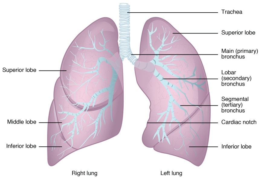
- The right lung is divided into three lobes, the superior lobe, middle lobe, inferior lobe. The left lung is divided into two lobes superior lobe and the inferior lobe.
- Each lobe of the lung is divided into small compartments called lobules, each lobule is covered by an elastic connective tissue that contains lymph vessels.
- Inside the lobule, the terminal bronchiole divides into the respiratory bronchiole which further divides into the alveolar duct. The alveolar duct is surrounded by alveoli and alveolar sacs.
Intercostal muscles:
- There are 11 pairs of intercostal muscles occupy the space between the 12 pairs of ribs. They are of two types:
- The external intercostal muscles.
- These extend downward and forward.
- When the intercostal muscles contract the ribs are pulled upwards towards the first rib which is non-moving or fixed. Due to the uplifting, the ribs expand in the forward direction leading to expansion of the chest cavity.
- The intercostal muscles are stimulated to contract by the intercostal nerves.
Diaphragm:
- The diaphragm is a dome-shaped muscular structure separating the thoracic and the abdominal cavities, it forms the door of the thoracic cavity and roof of the abdominal cavity.
- It consists of a central tender attached to the lower ribs and the sternum when the muscle of the diaphragm is operated the muscle fibers shorten and the central tendon is pulled rewards to the level of the 9th thoracic vertebrae enlarging the thoracic cavity.
- The diaphragm is supplied with the phrenic nerves.
Physiology of respiration:
- The physiology of respiration is divide into two phases External respiration: The exchange of gases (CO2 and Oy) between the alveoli of the lungs and blood in pulmonary capillaries is referred to as external respiration.
- The deoxygenated blood is pumped from the right ventricle to the pulmonary artery, which circulates the deoxygenated blood into the capillaries surrounding the alveoli. At the same time, the inspired atmospheric air enters the alveoli.
- In this particular situation the
The partial pressure of O2 in alveolar air is 105mm Hg and the partial pressure of O2 in capillary blood is 40mm Hg.
- Due to this difference in the partial pressure of oxygen, oxygen diffuses out from the alveolar air into the capillary blood. At the same time, there is a net diffusion of CO₂ from the capillary blood to the alveoli.
The factors affecting the partial pressure are:
- The partial pressure difference between the atmospheric gases.
- The thickness of the respiratory membrane.
- The surface area of the respiratory membrane.
- Solubility and molecular weight of the gases.
| QUESTION BANK 1. Write a neat labeled diagram explain the anatomy and physiology of lungs. (10M) |
Make sure you also check our other amazing Article on : Factors Affecting Renal Excretion of Drugs