
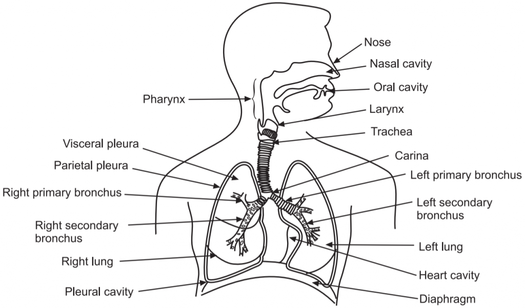
Breathing and Respiration: The human respiratory system consists of:
- The nose
- Pharynx (throat)
- Larynx (voice box)
- Trachea (windpipe)
- Bronchi
- Lungs.
Functions of Respiratory System:
- The respiratory system is responsible for gaseous exchange i.e. intake of O2 which is required by body cells and elimination of CO2, which is formed as a waste product by body cells.
- The respiratory system also helps to regulate blood pH.
- It filters inspired air, involved in the production of vocal sounds (phonation).
- The respiratory system also excretes small amounts of water and heat.
Mechanism of Breathing and Its Regulation
Respiration is the process of gaseous exchange in the body. This process is divided into three basic steps:
1. Breathing or pulmonary ventilation:
- Breathing is composed of two processes: inhalation and exhalation.
- The act of taking air in or inflow of air is called inhalation.
- The act of throwing air out or outflow of air is called exhalation.
- Thus, breathing involves the exchange of air between the atmosphere and the air of the lungs.
- During breathing, air flows between the atmosphere and the lungs because of alternating pressure differences created by the contraction and relaxation of respiratory muscles.
Mechanism of Breathing:
- Inhalation and exhalation are dependent on pressure differences in the lungs and atmosphere. The pressure difference is caused by changes in lung volume.
- The lungs must expand, for inhalation. Lung expansion increases lung volume and thus decreases the pressure in the lungs to below atmospheric pressure.
- The lungs expand with the help of contraction of the main muscles of inhalation, the diaphragm (the dome-shaped skeletal muscle that forms the floor of the thoracic cavity), and external intercostals (muscles present between ribs).
- Contraction of the diaphragm causes it to flatten, lowering its dome which increases the vertical diameter of the thoracic cavity. About 75% of the air enters the lungs during quiet breathing due to the contraction of the diaphragm.
- Contraction of external intercostals elevates the ribs. As a result, there is an increase in the anteroposterior and lateral diameters of the chest cavity. About 25% of the air enters the lungs during normal quiet breathing due to the contraction of external intercostals.
- As the volume of the lungs increases in this way, the pressure inside the lungs, called the alveolar (intrapulmonic) pressure, drops. A pressure difference is thus established between the atmosphere and the alveoli.
- Because air always flows from a region of higher pressure to a region of lower pressure, inhalation takes place. Air continues to flow into the lungs as long as a pressure difference exists.
- The process of exhalation is also dependent on the pressure gradient, but in this case, the gradient is in the opposite direction: The pressure in the lungs is greater than the pressure of the atmosphere.
- As no muscular contractions are involved, normal exhalation is a passive process.
- Exhalation starts when the inspiratory muscles relax. As the diaphragm relaxes, its dome moves superiorly owing to its elasticity.
- As the external intercostals relax, the ribs are depressed.
- These movements decrease the vertical, lateral, and anteroposterior diameters of the thoracic cavity, which decreases lung volume.
- In turn, the alveolar pressure increases. Air then flows from the area of higher pressure in the alveoli to the area of lower pressure in the atmosphere.
2. External (pulmonary) respiration or pulmonary gas exchange (Exchange of gases, transport of gases):
- It is the exchange of gases between the alveoli of the lungs and the blood.
- In this process, blood gains O2 from inhaled air and loses CO2 through exhaled air.
- External respiration in the lungs converts deoxygenated blood (depleted of some O2) coming from the right side of the heart into oxygenated blood (saturated with O2) that returns to the left side of the heart.
3. Internal (tissue) respiration or systemic gas exchange (Exchange of gases, transport of gases):
- Internal respiration is the exchange of gases between blood and tissue cells.
- During this process, blood loses O2, as it is transferred to cells and CO2 formed by cells is added to the blood.
- The exchange of O2 and CO2 between systemic capillaries and tissue cells is called internal respiration.
- As O2 leaves the bloodstream, oxygenated blood is converted into deoxygenated blood. Unlike external respiration, which occurs only in the lungs, internal respiration occurs in tissues throughout the body.
Regulation of Respiration
The respiratory center regulates the process of respiration. The respiratory center is a widely dispersed group of neurons, located bilaterally in the medulla oblongata and pons of the brain stem. It can be divided into three areas:
i. The medullary rhythmicity area in the medulla oblongata: It controls the basic rhythm of respiration.
ii. The pneumotaxic area in the pons: It co-ordinates the transition between inhalation and exhalation.
iii. The apneustic area, also in the pons: It co-ordinates the transition between inhalation and exhalation.
Respiratory Volumes
- The apparatus commonly used to measure the volume of air exchanged during breathing and the respiratory rate is a spirometer or respirometer. The record is called a spirogram.
- While at rest, a healthy adult averages 12 breaths a minute, with each inhalation and exhalation moving about 500 mL of air into and out of the lungs.
- The volume of one breath is called the tidal volume (TV).
- The minute ventilation (MV): the total volume of air inhaled and exhaled each minute is respiratory rate multiplied by tidal volume:
MV = 12 breaths/min. × 500 mL/breath
= 6 liters/min.
- Inspiratory reserve volume: By taking a very deep breath, we can inhale a good deal more than 500 mL. This additional inhaled air is called the inspiratory reserve volume. It is about 3100 mL in an average adult male and 1900 mL in an average adult female.
- Expiratory reserve volume: If we inhale normally and then exhale as forcibly as possible, we should be able to push out considerably more air in addition to the 500 mL of tidal volume. This is called expiratory reserve volume. It is 1200 mL in males and 700 mL in females.
- Inspiratory capacity is the sum of tidal volume and inspiratory reserve volume (500 mL 3100 mL = 3600 mL in males and 500 mL 1900 mL = 2400 mL in females).
- Functional residual capacity is the sum of residual volume and expiratory reserve volume (1200 mL 1200 mL = 2400 mL in males and 1100 mL 700 mL = 1800 mL in females).
- Vital capacity is the sum of inspiratory reserve volume, tidal volume, and expiratory reserve volume (4800 mL in males and 3100 mL in females).
- Total lung capacity is the sum of vital capacity and residual volume (4800 mL 1200 mL = 6000 mL in males and 3100 mL 1100 mL = 4200 mL in females).
Make sure you also check our other amazing article on : Body Fluids and Circulation