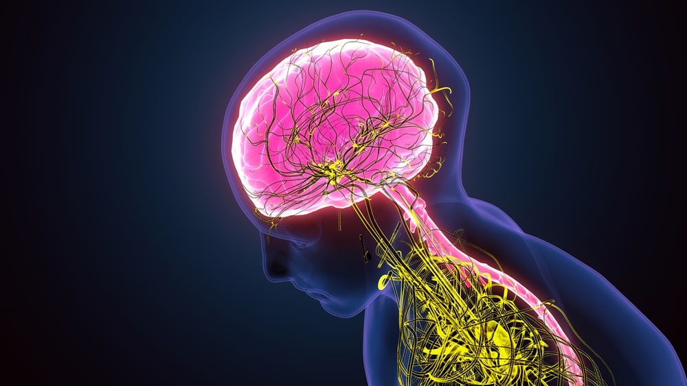
Neural Control and Coordination: The nervous system is a complicated, highly structured network of billions of cells called neurons and surrounding cells called neuroglia.
The nervous system is composed of the following parts:
- The brain
- Cranial nerves and their branches
- The spinal cord, spinal nerves, and their branches
- Ganglia
- Enteric plexuses
- Sensory receptors
Classification of Nervous System
Table of Contents
The nervous system can be classified into two main parts:
- The Central Nervous System (CNS): It consists of the brain and spinal cord.
- The Peripheral Nervous System (PNS): It consists of all nervous tissue outside the CNS.
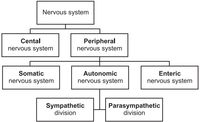
The peripheral nervous system is further divided into three parts:
- Somatic nervous system
- Autonomic nervous system (ANS): It is further divided into two divisions sympathetic and parasympathetic.
- Enteric nervous system
Structure of a Neuron
Nervous tissue consists of two types of cells Neurons and Neuroglia. Neurons are responsible for all functions of the nervous system like thinking, memory, sensing, etc. Neuroglia surrounds neurons and is responsible for supporting and providing nutrition to neurons.
Neurons consist of three parts: cell body, dendrites, and axon.
1. Cell body:
- It comprises a nucleus surrounded by cytoplasm.
- The cytoplasm of the cell body shows the presence of cellular organelles such as Golgi complex, lysosomes, mitochondria, free ribosomes, and prominent clusters of rough endoplasmic reticulum, termed Nissl bodies.
2. Dendrites:
- These are the processes that emerge from the cell body.
- Dendrites are generally tapering, small and extremely branched (tree-shaped).
- Dendrites play an important role in the transfer of signals from one neuron to other.
- Dendrites are receiving parts of a neuron.
- Their cytoplasm contains mitochondria and other organelles.
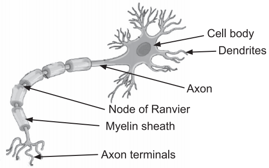
3. Axon:
- These are long, thin, cylindrical projections emerging out from the cell body of the neuron.
- Axons also have the presence of mitochondria, microtubules, and neurofibrils.
- The endoplasmic reticulum is absent in the axon.
- The cytoplasm of an axon is called axoplasm.
- Axoplasm is surrounded by a plasma membrane known as the axolemma.
- The end part of the axon is divided into many fine processes called axon terminals.
- Based on the presence of covering on the outside, the axons can be classified into two types:
- Myelinated axons: Axons surrounded by a multi-layered lipid and protein covering, called the myelin sheath, are called myelinated. The sheath electrically insulates the axon of a neuron and increases the speed of nerve impulse conduction.
- Unmyelinated: Axons without myelin sheath are called unmyelinated.
- The single axon of a neuron plays an important role in transferring nerve impulses to another neuron, a muscle fiber, or a gland cell.
- The location of communication between two neurons or between a neuron and an effector cell is called a synapse.
Generation and Conduction of Nerve Impulse
Nerve impulse-basic Concept:
- Neurons are excitable cells. Neurons are called excitable cells because they can respond to stimuli by generating electrical signals such as action potentials, also called nerve impulses i.e. electrical excitability.
- An action potential (nerve impulse) is an electrical signal which is generated by a neuron due to strong change in the environment (internal or external) and which travels along the surface of the membrane of a neuron.
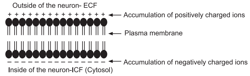
- The different concentrations of Na+ and K+ in the cytosol and extracellular fluid are crucial for the ability of neurons to generate electrical signals such as action potentials.
- As we know, proteins are present in the plasma membrane, some proteins called transmembrane proteins to act as ion channels, which regulate when ions can move in or out.
- Neuron shows a membrane potential, an electrical potential difference (voltage) across the membrane called the resting membrane potential.
- It exists because of the accumulation of negative ions in the cytosol along the inside of the membrane, and an equal accumulation of positive ions in the extracellular fluid along the outside surface of the membrane. This potential is measured in millivolts.
- The resting membrane potential of a neuron typically is –70 mV.
Generation of Action Potential:
- An action potential is generated due to the existence of a potential called resting membrane potential and the movement of ions (mainly Na+ and K+) across the membrane of neuronal cells.
- An action potential includes a sequence of quickly occurring actions across the neuronal membrane that decrease and reverse the membrane potential and then eventually restore it to the resting state.
- There are two phases in action potential:
- Depolarizing phase: In this phase, the negative membrane potential becomes less negative, reaches zero and then becomes positive.
- Repolarizing phase: In this phase, the membrane potential is restored to the resting state i.e. at –70 mV.
- Due to depolarization, when the potential across the membrane reaches a level called a threshold, an action potential is generated. Generally, the threshold potential in a neuron is -55 mV.
Process of Generation of Action Potential:
- The neuronal membrane is at resting state i.e. the potential across it is resting membrane potential. All ion channels are closed during this phase.
- A stimulus is responsible for the change in membrane potential. If due to any stimulus, when the membrane potential of the axon reaches a threshold, Na+ channels open and Na+ ions flow into the neuron from the extracellular fluid. This flow of Na+ ions increases charge inside the neuron which increases membrane potential. This is the depolarization phase.
- After depolarization, Na+ channels close and K+ channels open. K+ ions start moving outside the neuron to decrease the positivist caused due to excess entry of Na+ ions during the depolarization phase. This is called as repolarization phase. The outflow of K+ ions restores resting membrane potential.
All ion channels are closed.
Propagation of Action Potentials:
- Action potential spreads along the membrane. This mode of conduction is called propagation.
- The action potential travels down the axon as voltage-gated ion channels are opened by the spreading depolarization.
- There are two types of conductions: continuous and salutatory.
- In unmyelinated axons, this happens continuously because there are voltage-gated channels throughout the membrane.
- In myelinated axons, propagation is described as saltatory because voltage-gated channels are only found at the nodes of Ranvier and the electrical events seem to “jump” from one node to the next. Saltatory conduction is faster than continuous conduction.
- Thus, myelinated axons propagate their signals faster as compared to unmyelinated axons.
Structure of Brain and Spinal Cord
Brain
The brain is an important organ of the central nervous system. The brain is located in the skull. The brain is composed of four major parts:
- Cerebrum
- Cerebellum
- Diencephalon
- Brain stem
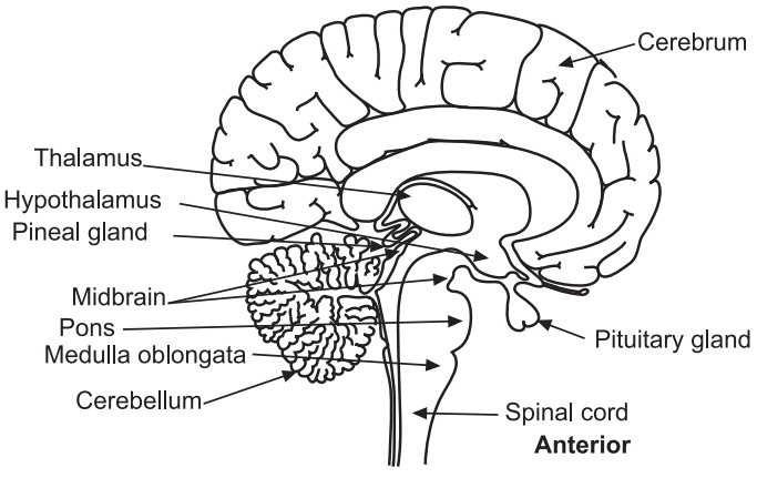
1. Cerebrum
- The cerebrum is the most important, largest, and uppermost part of the brain.
- The cerebrum consists of an outer cerebral cortex, an internal region of cerebral white matter, and gray matter nuclei deep within the white matter.
Cerebral Cortex
- The cerebral cortex is a region of gray matter that forms the outer border of the cerebrum.
- The folds present in the cortex are called gyri.
- The deepest grooves between folds are known as fissures; the shallower grooves between folds are termed sulci.
- The cerebrum consists of two hemispheres. The most prominent fissure, the longitudinal fissure, separates the cerebrum into right and left halves called cerebral hemispheres.
- The cerebral hemispheres are connected internally by the corpus callosum.
- Each cerebral hemisphere can be further subdivided into four lobes. The lobes are named after the bones that cover them: frontal, parietal, temporal, and occipital lobes.
- Functions
- The cerebrum is responsible for multiple activities like consciousness, thinking, memory, sensations, emotions, and willed movements.
Brain Stem
It is a slightly elongated posterior region of the brain which connects it to the spinal cord. The brain stem is divided into three parts: Medulla oblongata, Pons, and Midbrain.
Medulla Oblongata
It forms the inferior part of the brain stem. It is continuous with the superior part of the spinal cord.
Functions of Medulla Oblongata:
- The cardiovascular center controls the rate and force of the heartbeat and the diameter of blood vessels.
- The medullary rhythmicity area of the respiratory center regulates the basic rhythm of breathing.
- The vomiting center of the medulla causes vomiting.
- The deglutition center of the medulla promotes swallowing or deglutition of a mass of food that has moved from the oral cavity of the mouth into the pharynx (throat).
- Processes like sneezing, coughing and hiccupping are associated with the medulla oblongata.
2. Cerebellum
It is also called the little brain is located posterior to the brain stem.
- The cerebellum is posterior to the medulla and pons and inferior to the posterior portion of the cerebrum.
- The shape of the cerebellum looks like a butterfly.
- The central constricted area is called the vermis, and the lateral “wings” or lobes are the cerebellar hemispheres.
- Each hemisphere consists of lobes separated by deep and distinct fissures.
- Cerebellum has a highly folded surface that greatly increases the surface area of its outer gray matter cortex.
- The superficial layer of the cerebellum is called the cerebellar cortex.
- The cerebellum is separated from the cerebrum by a deep groove known as the transverse fissure, along with the tentorium cerebelli.
Functions of Cerebellum:
- The main function of the cerebellum is to regulate posture and balance of the body.
- It is also responsible for coordinating contractions of skeletal muscles. It produces smooth coordinated movements, maintains equilibrium, and sustains normal postures.
- The cerebellum may have a role in cognition and language processing.
Diencephalon:
It is a small region superior to the brain stem. It consists of three parts: Thalamus, Hypothalamus, Epithalamus.
(a) Hypothalamus: The hypothalamus is a small part of the diencephalon located inferior to the thalamus.
Functions of the Hypothalamus:
- Its control of activities of the autonomic nervous system.
- Hypothalamus is also called as body’s thermostat. It regulates body temperature.
- Hypothalamus is involved in the regulation of food intake. It comprises a feeding center, which promotes eating, and a satiety center, which causes a sensation of fullness and cessation of eating. It also contains a thirst center.
- Hypothalamus is connected to the pituitary gland, which is located inferior to it. Hypothalamus produces various hormones which affect the pituitary gland.
- Hypothalamus is the body’s internal biological clock.
- Hypothalamus is involved in the regulation of circadian rhythms.
- Hypothalamus regulates emotional and behavioral patterns.
Spinal Cord
- The spinal cord is located within the vertebral canal of the vertebral column.
- The spinal cord is roughly cylindrical and is flattened slightly anteriorly and posteriorly.
- Externally, the spinal cord shows two conspicuous enlargements – the superior enlargement is called the cervical enlargement and the inferior enlargement is called lumbar enlargement.
- Spinal nerves emerge at regular intervals from various regions of the spinal cord.
- Total 31 pairs of spinal nerves emerge out from the spinal cord.
- Spinal nerves are the routes of communication between the spinal cord and specific regions of the body.
Anatomy of the Spinal Cord:
- The transverse section of the spinal cord shows regions of white matter which surround an inner core of the gray matter.
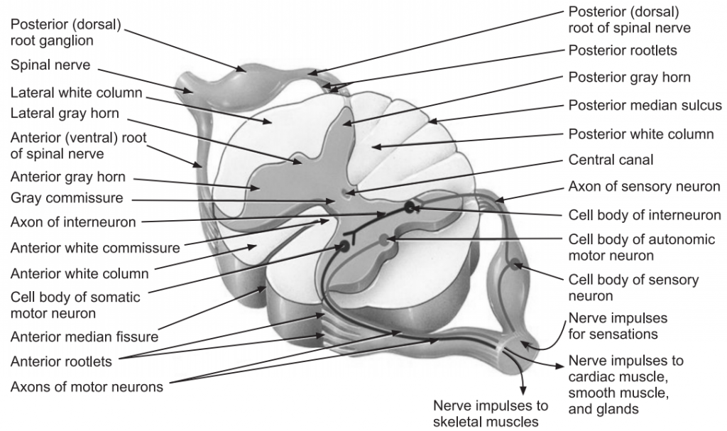
- Two grooves namely, anterior median fissure and posterior median sulcus penetrate the white matter of the spinal cord and divide it into right and left sides.
- The gray matter of the spinal cord is shaped like the letter H or a butterfly; the Gray matter of the spinal cord is composed of dendrites and cell bodies of neurons, unmyelinated axons, and neuroglia.
- The crossbar of the H is formed by the gray commissure.
- A small space called the central canal is located in the center of the gray commissure. The central canal is present the entire length of the spinal cord and is filled with cerebrospinal fluid.
- Anterior to the gray commissure is the anterior (ventral) white commissure, which connects the white matter of the right and left sides of the spinal cord.
- The gray matter on each side of the spinal cord is subdivided into regions called horns.
- The posterior (dorsal) gray horns contain cell bodies and axons of interneurons as well as axons of incoming sensory neurons.
- The anterior (ventral) gray horns contain somatic motor nuclei, which are clusters of cell bodies of somatic motor neurons.
- The anterior and posterior gray horns divide the white matter on each side into three broad areas called columns:
- anterior (ventral) white columns.
- posterior (dorsal) white columns.
- lateral white columns.
Make sure you also check our other amazing article on : Breathing and Respiration