Plant And Animal Tissues: An aggregation of similar cells or elements having the same function is known as tissue. The organs of plants or animals are composed of various tissues. Usually, in an organ, various tissues perform interrelated functions.
Plant Tissues
Depending upon their origin, structure and physiology, the plant tissues can be classified as follows: (I) Meristematic or embryonic tissues (II) Permanent tissues
(I) Meristematic Tissues: They have the following characters.
- The cells are small and isodiametric.
- The cells are very compact and without any intercellular spaces.
- The cells are thin-walled, normally cubical and with a prominent nucleus.
- Vacuoles in meristematic tissues are very small or even may be absent.
- Their cells can multiply.
- The newly formed cells due to enlargement and also due to morphological differentiation get transformed or changed into permanent tissue. Meristematic tissues in the plants are located at the growing points i.e. tips of the shoots and roots, between the bark and the wood of trees, in the bark themselves and also in any part of the plant wheresoever extensive growth of the plant takes place.
(II) Permanent Tissues: These exhibit the following characteristics:
- The cells vary in size, shape and nature of the protoplast.
- The cells may be living or dead.
- These are produced by meristematic cells.
- They do not have any capacity to multiply.
- They are not usually changed into other kinds of tissues and normally retain their structural physiological characters.
- They are composed of one or more types of cells.
Type of the cells, characters of the cell walls and the contents of the cells of the permanent tissues divide them into two categories namely, simple permanent tissue and complex permanent tissues.
(A) Simple Permanent Tissues:
As described above, this consists of only one type of cell forming a homogeneous structure. These are further sub-classified as (1) Epidermis, (2) Parenchyma, (3) Sclerenchyma, (4) Collenchyma, (5) Cork.
1. Epidermis: It is a type of simple permanent tissue forming the outermost layer of plant structure and normally only one cell thick and is made up of parenchyma. It occurs as the surface layer of leaves, flowers, fruits, seeds and younger parts of roots and stems.
Its function is to conserve the moisture supply to the inner tissues and offer protection to a certain extent against mechanical infection.
The outer walls of the epidermal cells are often thick and covered with a fatty substance cutin. The layer with cutin is called the cuticle. Minute epidermal openings, i.e. stomata, epidermal hairs i.e. trichomes are the other characters of the epidermis. Epidermal cells are colourless except when pigments are present.
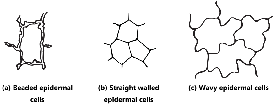
The structure of epidermal cells may vary, i.e.
- Straight walled polygonal, e.g. senna leaf
- Thick-walled beaded, e.g. Digitalis
- Wavy, e.g. Hyoscyamus
- Wavy and with striated cuticle, e.g. Belladonna
- Papillose, e.g. Stigma of pyrethrum flowers
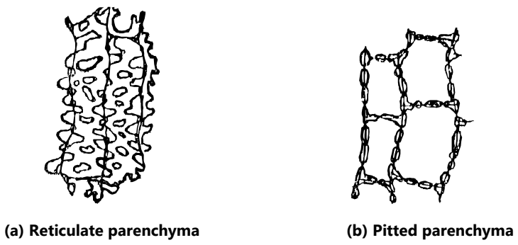
2. Parenchyma: This is another type of permanent tissue of almost all organs of higher plants.
Parenchymatous cells are thin-walled polyhedral in shape and with large central vacuole in many cases. Ageing results in the change in shape, wall thickening and the appearance of intercellular spaces.
The functions of parenchymatous cells are the storage of food and water, food manufacture, excretion, secretion and also assimilation. Parenchyma is a living tissue.
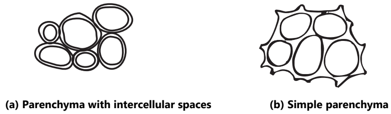
The secondary thickening may give rise to (1) Simple parenchyma (2) Parenchyma with intercellular spaces (3) Lignified parenchyma and (4) Reticulate parenchyma.
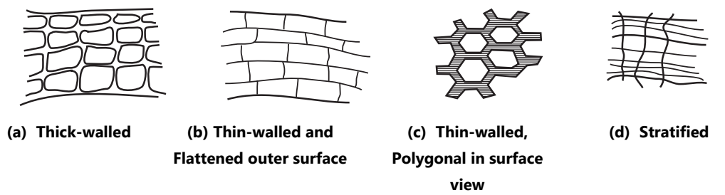
3. Sclerenchyma: It is a supporting tissue and is purely mechanical in its function. It is found in stems, roots, leaves and other parts along with the xylem. It is very hard and is responsible for the strengthening of the organs in which it is present.
It consists of two types of elements: (a) Stone cells (b) Fibres.
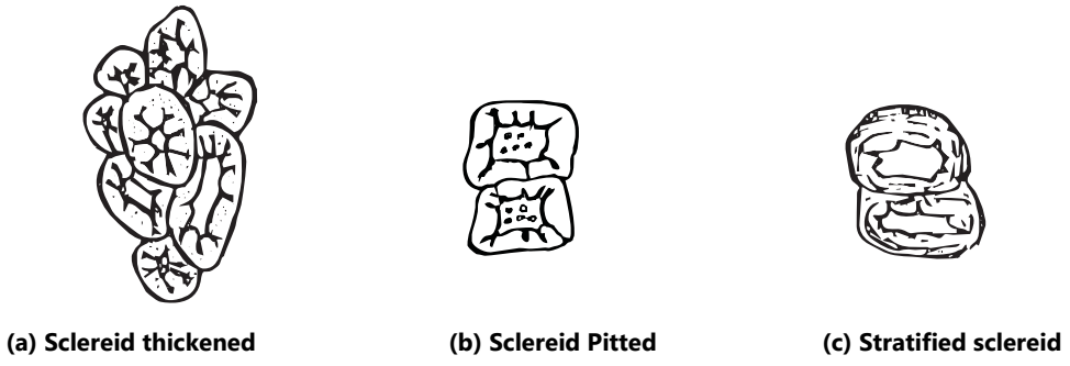
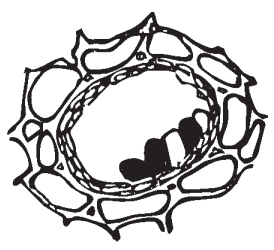
(a) Stone cells or Sclereids: These are more or less isodiametric, elongated or branched forms. Pitting or stratification may be also observed. They are found in groups, single or complete layers. Generally abundant in the cortex, phloem of stems and roots, maybe in seeds (cardamom) or flesh of fruits (pears).
(b) Fibres: Usually developed in bundles or layers. Usually thick-walled and have a narrow lumen and pointed ends. Walls of the fibres are usually lignified, but may also contain cellulose (ephedra contains non-lignified fibres, flax-fibres are also non-lignified). Depending upon the seat of their occurrence they are said to be pericyclic fibres, xylem fibres or phloem fibres. A sheath of crystals is formed around sclerenchyma and is a salient character for identification as in senna-leaf.
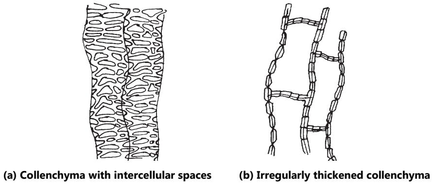
4. Collenchyma: It is the typical supporting tissue found underneath the epidermis of many stems, leaf stalks and leaf midribs. It is similar to parenchyma in structure except that the primary cell wall is thickened to give mechanical strength to plant parts. Collenchyma may or may not have intercellular spaces, may contain chloroplasts. Thickening may be stratified or unevenly distributed around the lumen.
5. Cork: It is a simple permanent tissue made up of compactly arranged approximately prismatic cells found in radial rows.
No intercellular spaces are present between them. It forms the bark of stems or roots. It is protective in function and guards against mechanical injury to inner bark or cambium, and also prevents loss of water. Protoplasm is the cork cell dies and hence matured cork cells are dead.
(B) Complex Permanent Tissues:
In this type of tissue, several types of cells are present which usually engage in a group of closely related activities. Following are the various types of complex tissues found in plants.
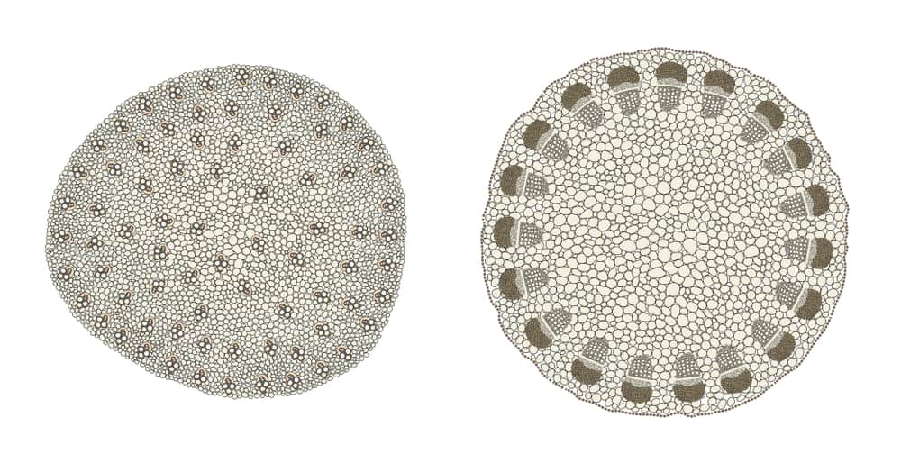
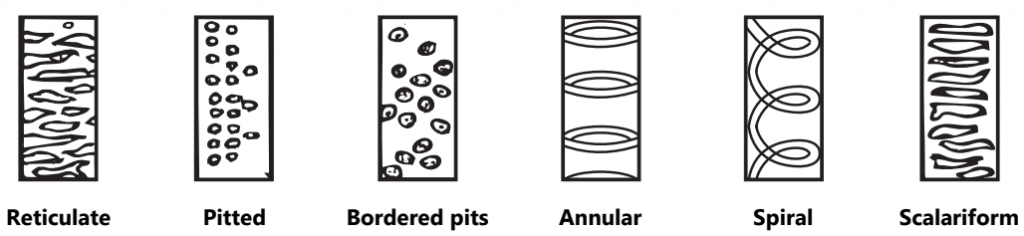
(a) Xylem: It is the main water-conducting tissue of the plant always associated with the phloem in the vascular tissues. The components of the xylem are parenchyma, fibres, and tracheary elements. Tracheary elements namely tracheids and vessel members are highly specialized cells elongated in structure and non-living in nature.
The thickening of xylem vessels is lignified. Tracheids are closed (imperforate) while the vessels are open at both the ends and form the tube. The thickening to the xylem elements may be annular, spiral, reticulate, pitted, border pitted and scalariform.
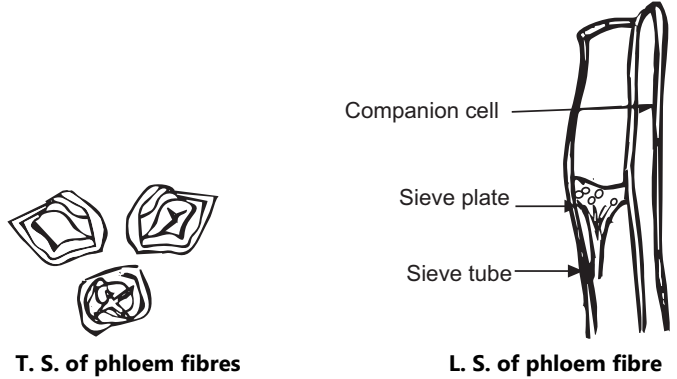
(b) Phloem: It is mainly responsible for the transport of food manufactured in the leaves down to stem and roots. Other types of cells constituting the phloem are phloem parenchyma, sieve tubes, companion cells and phloem fibres. Sieve tubes contain living protoplasm. The end walls of sieve tubes contain circular perforations. There are perforations in between sieve tubes and companion cells. Phloem parenchyma, ray cells store the food while phloem fibres are meant for strength and support.
(c) Secretory Structure: These are important features of certain plants containing secretions. They may be internal or external. These structures may be single-celled or multicellular. The oil gland and resin cells are unicellular, while the ducts or glands are multicellular.
They may get formed by splitting of cells i.e. schizogenous or by the breakdown of a group of cells i.e. Lysigenous or by a combination of the former and the latter i.e. schizolysigenous. The secretions may be tannins, oils (volatile or fixed) mucilage and crystals.
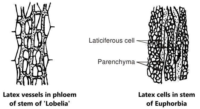
Laticiferous tissue: These are also known as milk tubes. This tissue is found in plants belonging to certain families. They may be in the form of vessels or cells. The white juice which they contain may be thick, or watery, or maybe white (papaya, euphorbia) or coloured (orange in chenopodium and poppy).
These vessels are not only branched but reunited to form a network. Their function is not known. Latex contains several chemicals like alkaloids, enzymes, proteins etc.
Conducting Tissue System:
Vascular elements meant for the transport of the food material are placed vertically. Vascular elements are continuous from the root to the leaf and are grouped in the bundle; such a structure is known as a vascular bundle which bundle consisting of xylem and phloem.
Following are the various types of vascular bundles found in the plants.
1. Radial: These are the vascular bundles comprising xylem and phloem only. They are arranged on alternate radii and hence known as radial. In roots, the xylem is exarch with the proto-xylem pointing towards the periphery and the metaxylem towards the centre.
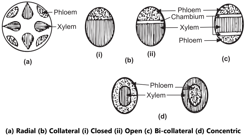
2. Collateral: In this case, the xylem and phloem are placed on the same radius xylem lies towards the centre and the phloem towards the periphery. There are two types of these vascular bundles. (a) Open collateral: When cambium is placed in between xylem and phloem and occur in dicotyledonous plants. (b) Closed collateral: When no cambium is present in between xylem and phloem this type is found in monocotyledonous plants.
3. Bi-collateral: In this case, the xylem lies in the centre with cambium and phloem on either side along the same radius. Bicollateral vascular bundles are found in the Cucurbitaceae family.
4. Concentric: Vascular bundles wherein there is no cambium but (a) either the xylem lies in the centre surrounded by phloem (Centroxylic) or (b) Phloem in the centre surrounded by xylem (Centrophloic). The first type is reported in ferns while the second is reported in Acorus (sweet flag).
Animal Tissues
The outer nuclear membrane bears ribosomes which may continue with the membrane of the endoplasmic reticulum.
Mitochondria:
Within the cytoplasm, there are numerous double-layered elongated bodies present called mitochondria. It is composed largely of proteins and lipids. Each mitochondrion is composed of tubular or paired lamellae called cristae. It is the site for carbohydrate and lipid (fat) metabolism i.e., respiration.
Golgi Apparatus:
The Golgi apparatus (body) is compact and consists of parallel membrane plates and tubules. It is the site for enzyme secretion.
Endoplasmic Reticulum and Ribosomes:
Within the cytoplasm of the cell is an extensive network of membranes arranged in plates and tubules, collectively known as the endoplasmic reticulum. It is the site for biochemical synthesis and intracellular transport of molecules.
The endoplasmic reticulum present is attached to the nuclear membrane and the plasma membrane. On the membrane of the endoplasmic reticulum, there are small round bodies present called ribosomes. It is the site for protein synthesis. The endoplasmic reticulum with the ribosomes is called the rough endoplasmic reticulum, while devoid of ribosomes it is called the smooth endoplasmic reticulum.
Lysosomes:
The lysosomes are small vesicular structures containing homogeneous fluid. It is composed of a single layer and contains digestive enzymes for intracellular digestion.
Microtubules:
These are tubular structures composed of a globular protein and held for intracellular transport.
Microfilaments:
These are protein filaments in the cytoplasm meant for contraction and motility of the cell.
The animal cell is responsible to perform functions like motility, digestion, metabolism, growth, reproduction and irritability.
Animal Tissues:
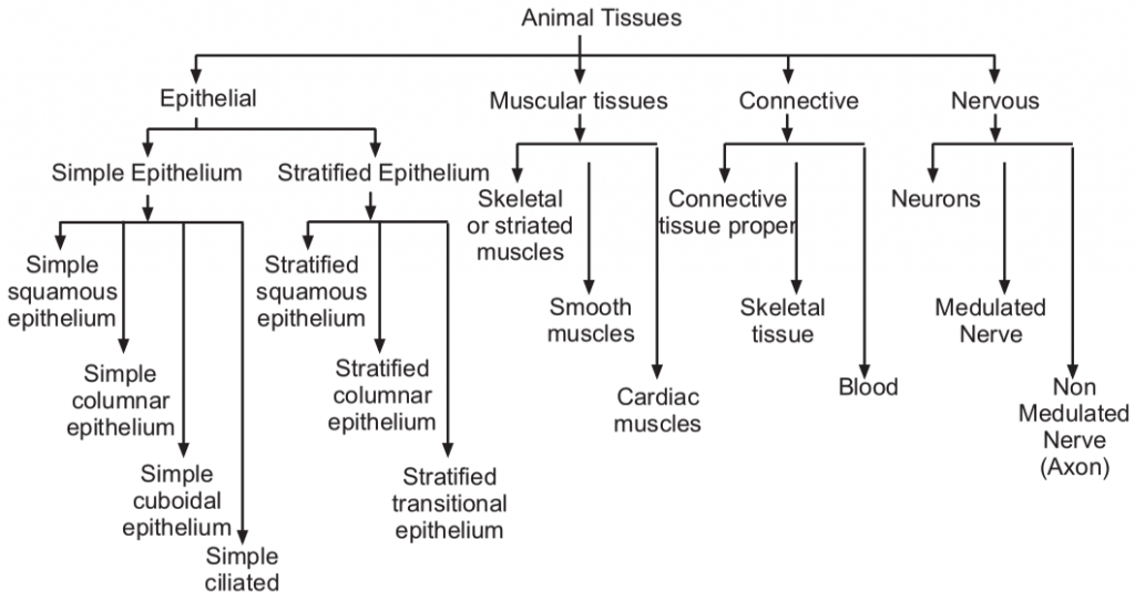
The animal body is composed of cells that are grouped to form tissue. An organ of the body consists of various tissues of common function. The tissues may be simple or compound. A simple tissue is a combination of similar cells while different kinds of cells together form a compound tissue. Simple tissues can be broadly classified into four groups 1. Epithelial, 2. Muscular, 3. Connective, and 4. Nervous.
1. Epithelial Tissues: Epithelial tissue is a group of cells present on the free surface of organs or lining of cavities. The cells are compound, arranged with little intercellular space. The functions of epithelial tissues are protection, absorption, secretion, excretion, reproduction etc.
Epithelia are classified according to their shape and the arrangement of cells.
These are grouped into simple and stratified epithelia. The simple epithelia are (a) Squamous, (b) Columnar, (c) Cuboidal and (d) Ciliated. The stratified epithelia are (i) stratified squamous, (ii) stratified columnar and (iii) stratified transitional.,
A. Simple Epithelium:
(a) Simple Squamous Epithelium: It consists of a single layer of flattened cells. The nucleus is oval and present in the centre of the cell. This type of epithelium can be obtained from the skin of the frog or Bowman’s capsule and Henle’s loop of mammal. (Fig.12)
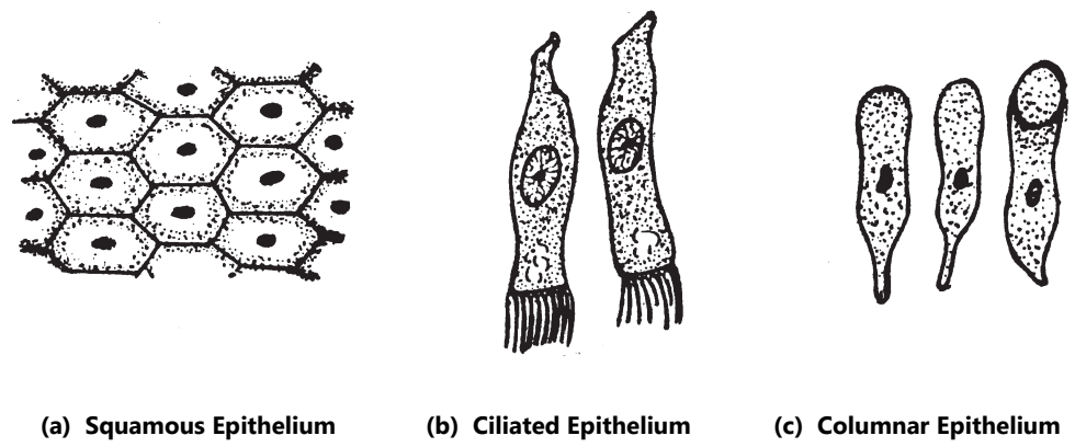
(b) Simple Columnar Epithelium: It forms a single layer of elongated cells arranged side by side like columns. It can be obtained from the lining of the intestine of a frog or the inner lining of the stomach, small intestines in mammals. The elongated cells have free broad ends while the narrow ends are arranged side by side with the nucleus inside. Some cells secrete slime or mucous in vacuoles kept towards their broad ends. (Fig.12)
(c) Simple Cuboidal Epithelium: It forms a single layer of cubical or polygonal cells with equal dimensions on all sides. The nucleus is round and centrally placed. These epithelia are found in ducts of various glands and germinal epithelium.
(d) Simple Ciliated Epithelium: The cells are elongated and based on the basement membrane. They are generally of the columnar type. The nucleus has no particular place in the cell. These are found in the buccal cavity of frogs and trachea in mammals. (Fig.12).
B. Stratified Epithelium:
It has two or more layers of the simple epithelium.
(i) Stratified Squamous Epithelium: It consists of three layers of cells. The first layer consists of flattened and fusiform cells, the second polygonal cells and the third basal cuboidal cells. The outermost layer is cornified. The cells of the basal layer consist of spherical nuclei. This type of epithelium is found in the oesophagus and epidermis.
(ii) Stratified Columnar Epithelium: It also consists of three layers of cells. The superficial layer consists of elongated columnar type. The middle layer has polygonal cells and the basal layer has cuboidal cells. The superficial layer cells consist of oval nuclei while other layer cells consist of spherical nuclei. These are located in the urethra of mammals.
(iii) Stratified Transitional Epithelium: Here the superficial layer consists of dome-shaped cells, the middle layer with club-shaped cells and the lower basal layers have cuboidal cells. The nucleus is round or oval. These epithelia are found in the urinary bladder.
Functions of Epithelium:
Epithelium covers the free surface of the organ or body. It protects the body from the loss of fluid. It helps in the functions of absorption excretion, secretion etc.
2. Muscular Tissues:
These consist of long elongated muscle fibres held together by connective tissues. It shows a high degree of elasticity i.e. contraction and expansion of muscles, due to which it performs various kinds of movements. Muscular tissues can be divided into striped or striated and unstriated or smooth muscles.
The striated muscles are voluntary in their function i.e. they are controlled by the will, while, the unstriated muscles are involuntary i.e. they perform the functions without any order of the will. Striated muscles are found in various parts of the body, while unstriated muscles are located only in a few organs like the stomach, intestine and urinary bladder.
The muscular tissue can be divided into three groups: (a) Striated or skeletal muscles (b) Smooth or visceral or non-striated muscles and (c) Cardiac muscles.
(a) Striated Muscles: These consists of long elongated and multinucleated muscle fibres. These are attached to bones and are voluntary in their functions. These help in skeletal movement. These show alternate light and dark bands and hence are called striated. These have great power of contraction and are found in hands, legs, attached to bones, hence called skeletal muscles. [Fig.13 (a)].
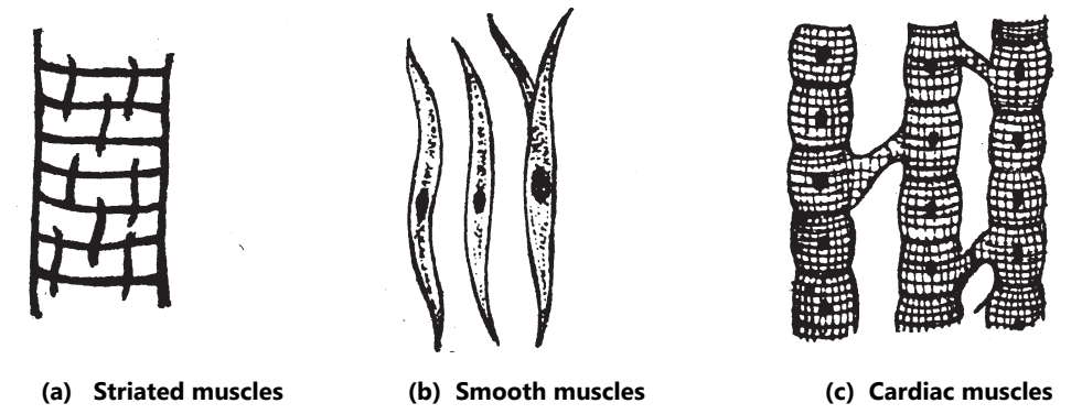
(b) Smooth Muscles: The fibres are long but are shorter than the striated muscles. These are spindle-shaped with a nucleus in the centre. Smooth muscles are involuntary in their function. They are composed of longitudinal microfibrils with no striations and hence are called non-striated or fibrous muscles. There is no sarcolemma but the cells are joined together by connective tissues. [Fig.13 (b)].
Smooth muscles contract slowly. These are found in the stomach, intestine, urinary bladder, uterus blood vessels and trachea.
(c) Cardiac Muscles: Cardiac muscles are found in the walls of the heart and are involuntary in their function. The fibres are long cylindrical and branched repeatedly to form a network. Each fibre has a centrally located nucleus, surrounded by sarcoplasm. These muscles help in the rhythmic contraction of the heart. [Fig.13 (c)].
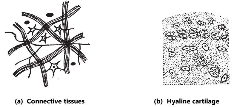
3. Connective Tissues:
These are loosely packed supporting cells. Connective tissues are present in the intercellular substances or ground substances of the matrix. The various types of connective tissues perform different functions. Some connective tissues connect the organ while others are packing and skeletal in their function.
Connective tissues can be divided into three groups: (i) Connective tissue proper; (ii) Supporting or skeletal tissue; and (iii) Fluid or blood tissue.
(i) Connective Tissue Proper: These tissues connect organs or parts. It consists of areolar and fibrous tissues. Areolar tissue is present beneath the skin and matrix. In the matrix, two kinds of fibres are present called white fibres and yellow or elastic fibres. The white fibres are wavy, unbranched and found in bundles. The yellow fibres are elastic, single, straight and branched. The areolar connective tissues in nerve fibres, muscle fibres, connect the skin with the body wall and is surrounded by blood vessels. These are found in the heart and blood vessels. [Fig.14 (a)].
Any excess amount of fat is deposited in the areolar tissues present beneath the skin. These fatty tissues are known as adipose tissues. The fibrous tissues connect the muscles with the bone.
(ii) Supporting or Skeletal Tissue: It consists of bone, cartilage and hyaline cartilage. The cartilage is found on the tip of the bones of girdles and limbs. These are also found in the trachea and the larynx. The cartilage with yellow elastic fibres in the matrix is called cartilage and is found in the external ear and the nose. The hyaline cartilage consists of many elastic fibres (Fig.14 (b)). Most of the endoskeleton is made of bones. The bone comprises the hardest tissues.
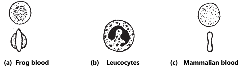
(iii) Fluid or Blood Tissue: Blood is called fluid conductive tissue. Blood is present in vessels, like the arteries and the veins. The blood consists of plasma in which corpuscles are suspended. These are erythrocytes (R.B.C.) and leucocytes (W.B.C.). The plasma corresponds to the matrix and the corpuscles, to the cells. The corpuscles are round and nucleated in frogs and non-nucleated in a mammal. The leucocytes are irregular in shape with nuclei inside. (Fig.15).
4. Nervous Tissue:
Nervous tissues are formed by nerve cells and nerve fibres. These can be divided into (a) nerve cells or neurons, (b) medullated nerves and (c) non-medullated nerves.
(a) Neurons: The neurons are found in the brain, spinal cord and nerve ganglia. A typical neuron has a large cell body containing a nucleus. From the cell body, several fine nerves develop called dendrites. A single long nerve arising from the cell body is called an axon. The axon conducts impulses away from the cell body while dendrites towards the cell body. The axon is covered by a sheath called the medullary sheath. The medullary sheath is not continuous but is broken in the middle. The breaks are called nodes of Ranvier. (Fig.16).
(b) The medullated nerve is an axon covered by a medullated sheath, which is the sheath covered by a layer called neurolemma. It is a single layer of nucleated cells and is known as a white nerve fibre. At intervals, the sheath breaks to form nodes of Ranvier.
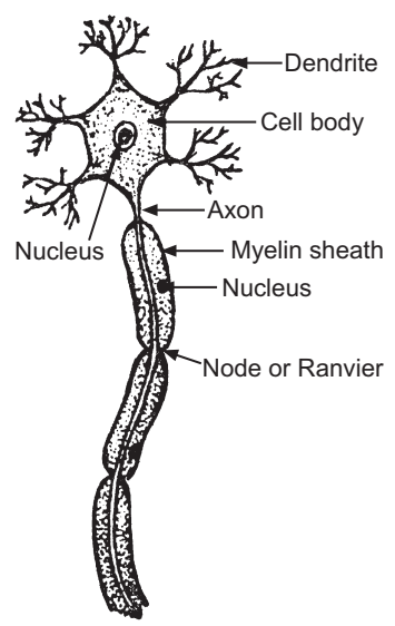
(c) The axon without medullated sheath is called a non-medullated nerve where the axon is directly covered by neurolemma. These are called grey nerve fibres. These nerve tissues receive and convey impulses of stimuli.
Ergastic Substances in Animal Cells:
As stated earlier the non-living substances of the cells are known as ergastic substances. Like plants, they are also produced by animals. They may be of three types: (1) Reserve foods; (2) Excretory products; (3) Secretory products.
The animal cell differs from plant cell to a great extent and hence the cell inclusions of animal cells vary. Following are the important differences between a plant cell and an animal cell.
| Animal Cell | Plant Cell |
| 1. Cell wall is absent. | 1. Cell wall is present. |
| 2. Plasma-membrane is the outermost boundary of the cell. | 2. Cell wall is the outermost boundary of the cell. |
| 3. Plastids are absent. | 3. Plastids are present. |
| 4. Lysosomes are present. | 4. Lysosomes are usually absent. |
| 5. Pair of centrioles is present. | 5. Centrioles are absent. |
| 6. Vacuoles are absent if observed are many and small. | 6. Large central vacuole is present. |
| 7. Phagocytosis or Pinocytosis is observed. | 7. Phagocytosis or Pinocytosis is not observed. |
| 8. Golgi apparatus is present with specific polarity. | 8. Golgi apparatus are scattered in the cytoplasm. |
Make sure you also check our other amazing article on : Plant Growth and Development