Principle and Procedure of Gram Staining: Gram staining is one of the most fundamental and widely used techniques for the differentiation and identification of bacteria. This differential staining technique was discovered by Dr. Christian Gram in 1884. It not only reveals the size and shape of bacteria but is also used to differentiate bacteria into Gram-positive and Gram-negative cells. Hence, it is known as differential staining.
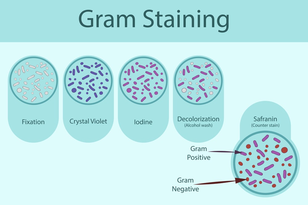
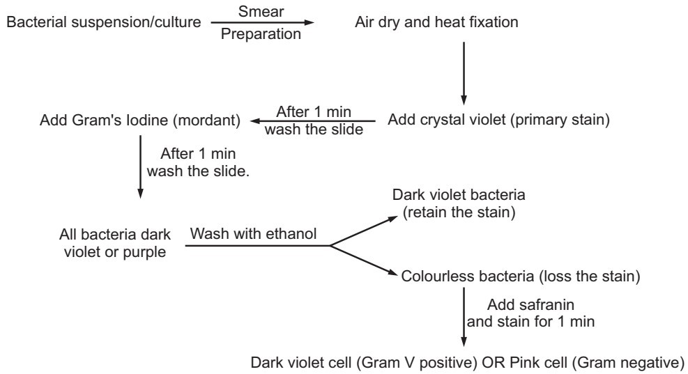
In this staining, the cells are stained with a basic stain (crystal violet) and treated with an iodine-potassium iodide mixture to fix the stain. Then, the stain is washed with alcohol or acetone (decolorizer) and counterstained with a polar dye of a different color (e.g. safranin). All bacteria take up the initial violet stain but only Gram-positive cells retain it during the subsequent steps (Bacillus anthracis, Clostridium tetani, Staphylococcus aureus, etc.). Gram-negative bacteria are easily decolorized and take up the counterstain and appear red (Escherichia coli, Salmonella typhi, Vibrio cholerae, Klebsiella pneumonia, etc.).
In Gram staining, four different reagents or stains are used.
1. Primary stain (crystal violet): The crystal violet stain is used first and stains all cells deep violet in color.
2. Mordant (Gram’s iodine): A mordant may be defined as ‘any substance that forms an insoluble compound with stain and serves to fix the color to bacterial cell’. It should be noted that mordant is not a stain. It leads to the form of crystal violent iodine – magnesium ribonucleate [CV-I-Mg ribonucleate] complex in Gram-positive bacteria. This complex is not formed in Gram-negative bacteria, as Mg-ribonucleate is absent in the cell wall. Hence, only the CV-I complex is formed in Gram-negative bacteria.
3. Decolourising agent (ethyl alcohol, 95%): The cell wall of Gram-positive cells contains less lipid. On application of decolorizing agents like alcohol or acetone, shrinkage of the cell wall takes place due to dehydration and decreases the permeability of the CV-I-Mg ribonucleate complex. Thus, the complex is retained in the cell and hence cell is stained deep violet. On the other hand, the treatment of decolorizing agent extracts lipids from the cell wall of a Gram-negative cell and there is an increase in the permeability property of the cell wall. Due to this, CV-I complex is extracted and cells get decolorized (lose violet color).
4. Counterstain (safranin): This is the final reagent to stain red, those cells that have been previously decolorized. Since only Gram-negative cells are decorated, they may now absorb the counter stain. Gram-positive cells retain the violet color of the primary stain. The procedure of staining is given in Fig.1.
Make sure you also check our other amazing Article on : Types of Electron Microscopes