
Skin Color Measurement: As one of the most conspicuous human polytypic variations, skin color has probably attracted more scholarly attention than any other aspect of human variability. Skin color has served as a primary feature in most systems of racial classification. The biological determinants of skin Color include the pigments carotene, hemoglobin, and melanin. Carotene, the least common skin pigment results in a yellowing of skin –results primarily from the over-consumption of carotene-containing foods (like carrots) –This pigment is significant almost exclusively in pathological or abnormal skin Coloration. Hemoglobin is the complex molecule responsible for the transport of oxygen throughout our bodies – It is the primary protein constituent of red blood cells. Oxygenated hemoglobin has a reddish hue and produces a pinkish tint to lightly pigmented skin, whereas deoxygenated hemoglobin has a purplish color and produces a bluish tint to lightly pigmented skin that is characteristic of oxygen deprivation and suffocation. The primary determinant of variability in human skin color is the amount, density, and distribution of the pigment melanin. Melanin has a dark brown/purple/black color that is intensified by denser compaction of the melanin granules in the cells of the upper layers of the skin.
Epidermis
Table of Contents
The epidermis is the superficial layer of the skin and consists of stratified squamous epithelium that is keratinized (containing the protein keratin). It forms the tough outer covering of our body allowing us to withstand physical, chemical, and thermal insults. Because the epidermis is avascular, it relies on the underlying dermis for nutrient and gas exchange. Four basic cells are forming the epidermis: keratinocytes, melanocytes, dendritic (Langerhans) cells, and tactile (Merkel) cells. The epidermis is composed of five layers in thick skin, the skin located at the soles of the feet and palms of the hands. The layers include: stratum corneum, stratum lucidum, stratum granulosum, stratum spinosum, and stratum basale (Fig.1). The skin located throughout the rest of the body is categorized as thin skin. It lacks the stratum lucidum seen in thick skin.
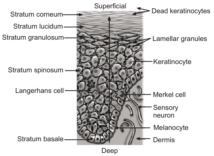
The stratum basale consists of columnar cells, the keratinocytes with about 10% of the cells comprising melanocytes. This is the germinal level of the skin which gives rise to the outer layers of cells and the melanin granules that pigment them. This is also where 90% of Vitamin D synthesis takes place. Melanocytes synthesize melanin which is combined into granules and injected into the surrounding keratinocytes. Melanocytes synthesize melanin which is combined into granules and injected into specialized cellular vesicles or organelles called melanosomes (measuring up to 500 nm in diameter). In dark skin, melanosomes accumulate eumelanin [which gives skin a brown color (tan)] and remain as single particles, while in lighter skins they contain increasing amounts of phaeomelanin [which gives skin a red color (burn)] and cluster in membrane-bound organelles, lysosomes. Skin color is primarily determined by genetic inheritance but exposure to sunlight also alters skin color.
Skin type
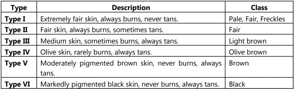
Skin type is a classification based on the skin’s reaction to exposure to the sun’s UV radiation, after a period of non-exposure (e.g. sun exposure at the beginning of summer, when the skin has not been exposed during winter). The skin’s reaction is measured in terms of burning and tanning. The Fitzpatrick system of classifying skin type is most commonly used in the assessment of skin cancer risk.
Table.1: The Fitzpatrick skin types
Measurement of Skin Color
By the later half of the nineteenth century, while anthropologists still had no clear idea of the underlying causes of pigmentation, they began to devise measurement techniques to use skin color in racial classification. Broca established a 34 tone scale, which was simplified by his student Topinard. These techniques were used into the 20th century until the introduction of the reflectance spectrophotometer in the early 1950s.
In dermatologic, practice, and clinical research, visual cues such as Color are of primary importance for the accurate diagnosis and grading of skin lesions. Quantification of erythema and pigmentation is important for in vivo assessment of skin reactions to external stimuli such as ultraviolet radiation. Measurement of lesion color is also useful for quantitative evaluation of the efficacy of therapies for skin lesions. However, visual inspection is a matter of perception and subjective interpretation and is hardly quantifiable. This is because there is such a wide variety of ways to express a color which makes describing a color or color difference extremely difficult and vague. Despite the ability of human eyes to recognize up to millions of colors, we are unable to precisely quantify our color perception without instrumental means. Therefore, there is a need for objective, scientific-based and non-invasive quantification of skin color or the extent of erythema and pigmentation. Reflectance spectrophotometers are widely used for the determination of skin color.
Reflectance Spectrophotometer
A Reflectance spectrophotometer emits light of a specific wavelength, using a filter, and measures the intensity of light reflected by the skin. Two types of skin reflectance instruments are available nowadays for the determination of skin color: a tristimulus Colorimeter (Chromameter from Minolta) using the CIE L*a*b* color system and the narrow-band simple reflectance meters (Derma Spectrometer from Cortex and Mexameter from Courage-Khazaka) using the erythema/melanin indices.
Chromameter
Chromameters or spectrophotometers are usually used for the observation of skin color. To quantify the changes in skin color, the CIE L*a*b* color system is often used. The L*a*b* parameters provide a measure of the perception of skin Color and can therefore emulate how the dermatologist or the average person perceives skin tones. The L*, b* values are often used to evaluate the amount of epidermal melanin, while the a* value is used to evaluate the amount of erythema (also referred to as hemoglobin) in the superficial plexus. In an attempt to quantitate skin pigmentation, the ‘Individual Typology Angle (ITA)’ or ‘Alpha Characteristic Angle (ACA)’ have been proposed.
Erythema and melanin indices are the indicators that quantify the intensity of erythema and pigmentation, respectively. These indices are derived from reflectance data of the skin at selected spectral bands. Unlike color coordinates, these indices are designed to show quantities that correlate linearly with the amounts of hemoglobin and melanin in the skin. Therefore, they can be handled as genuine physical quantities. The main advantage of using Chromameter with Skin Analysis Software CM-SA over the conventional narrow-band simple reflectance meters is its capability to provide both Colorimetric data (i.e., L*a*b*), as well as, the melanin and hemoglobin indices.
The CR-400 Chroma Meter is a handheld, portable measurement instrument designed to evaluate the color of objects, particularly with smoother surface conditions or minimal color variation. Through standard or customized evaluation formulae, this high accuracy, reliable Colorimeter helps users to control the color quality, consistency, and appearance of their samples in a more efficient, streamlined process internally and throughout the supply chain. It accurately identifies color characteristics in objects, determines color differences between objects, and provides pass/fails assessments to immediately determine if the sample meets the defined standard. This makes the CR-400 ideal for color inspections of food, building material, plastic, and dermatological applications within quality control, quality assurance, and R&D fields.
The CR-400 Chroma Meter is compatible with an optional data processor to print results on-site or SpectraMagic NX software to record measurements and provide a more comprehensive color analysis.
Mexameter®
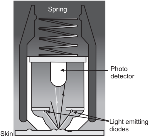
The Mexameter is a very easy, quick, and economical tool to measure the two components, mainly responsible for the color of the skin: melanin and hemoglobin (erythema) by reflectance.
The Measuring Principle
The measurement is based on absorption/reflection (Fig.2). The probe of the Mexameter emits three specific light wavelengths. A receiver measures the light reflected by the skin. As the quantity of emitted light is defined, the quantity of light absorbed by the skin can be calculated. The melanin is measured by specific wavelengths chosen to correspond to different absorption rates by the pigments. For the erythema measurement, specific wavelengths are also used, corresponding to the spectral absorption peak of hemoglobin, and to avoid other color influences (e. g. bilirubin).
Advantages:
- Very quick and easy measurement (1 sec for the two results: melanin index and erythema index).
- Continuous measurements over a longer period can optionally be performed.
- The highly sensitive measurement gives values on a broad scale (0-999) for melanin and erythema so that even the smallest changes in color become traceable.
- The probe is small and lightweight for easy handling and measurement on all body sites.
- Spring in the probe head ensures constant pressure on the skin enabling exact, reproducible measurements.
- The accuracy of the Mexameter® probe can be checked easily anytime.
- The probe head can easily be cleaned after each measurement.
Applications:
There are many fields of application where changes in skin color are of interest.
- In all important dermatological basic research and cosmetic application fields.
- It is indispensable in efficacy testing and claim support for cosmetics and pharmaceuticals (especially sunscreen and skin whitening products, skin-soothing).
- In occupational health, skin irritation (erythema value) is of special interest to show the necessity of skin protection measures.
Derma Spectrometer
Derma spectrometer provides a new and innovative approach to color measurement read-out of the erythema and melanin indices based on the absorbance characteristics of the human skin. It is fully handheld, battery-powered, and microprocessor-controlled to provide fast easy, and safe operation. Its fully handheld and lightweight design takes advantage of the latest development in color sensing technology, and the cable-connected color sensing probe offers the highest degree of freedom and flexibility in operation. Further, a special lens arrangement focuses on the target area and highly reduces the influence of ambient light. The instrument provides an easy selection of different color systems, and calibration by using the supplied calibrator is done in seconds.
Make sure you also check our other amazing Article on : Tewameter