Structure and Functions of Cell
Table of Contents
In 1838, Schleiden and Schwann put forward the cell theory stating that the plant or animal body is ultimately made up of minute cells and concluded that the cell is the structural unit of life.
The living organisms are of two types, either unicellular or multicellular.
In multicellular organisms, the life activities are performed by co-ordination of several organs, these organs are made up of tissues, while the tissues in turn are aggregates of similar cells. Thus, a cell is a structural unit of life. While in unicellular organisms all activities are performed by the same cell.
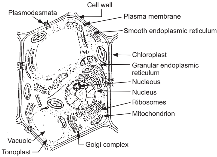
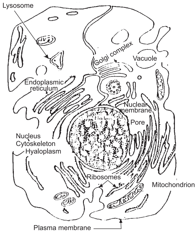
Structure of Cell
The cell has a definite shape of its type with a few exceptions like an amoeba, leucocytes, etc. Various shapes like oval, spherical, polyhedral, columnar, cylindrical, stellate and several others are found in the plant as well as in animal cells. Normally the size may vary from 0.5 to 20 µ and very exceptionally up to 200 – 300 mm as in the case of plant fibres and latex cells.
There are two types of cells. These are:
- Prokaryotic
- Eukaryotic
Prokaryotic cells are characterized by the absence of a true nucleus, obviously the nucleolus, the nuclear membrane is missing, DNA is without protein sheath and the nuclear matter is in direct contact with the cytoplasm. The ribosomes are scattered in the matrix while other organelles and endoplasmic reticulum are missing in prokaryotic cells. Respiratory enzymes and photosynthetic pigments are present. Meiosis and mitosis are not observed in this type of cell.
The primitive type of organisms like blue-green algae, bacteria, represents this type of cell. Eukaryotic cells possess a well-marked true nucleus, with a nuclear membrane, while DNA is covered with protein sheath. There is a distinct nucleolus in the nucleus, while plastids and mitochondria are represented in the cytoplasm. The other important and major component of the cytoplasm is the endoplasmic reticulum associated with ribosomes. In eukaryotic cells, the cell wall is made up of cellulose. Meiosis and mitosis are observed in these types of cells. All plant and animal cells belong to a eukaryotic type of cells.
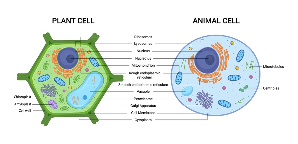
Difference between plant and animal cells:
Before knowing the structure and functions of various cell organelles, one must know the structural difference between a plant cell and an animal cell.
Table.1: Difference between plant and animal cell
| Animal Cell | Plant Cell |
| 1. Cell wall is absent. | 1. Cell wall is present. |
| 2. Plasma membrane is the outermost boundary of the cell. | 2. Cell wall is the outermost boundary of the cell. |
| 3. Plastids are absent. | 3. Plastids are present. |
| 4. Lysosomes are present. | 4. Lysosomes are usually absent. |
| 5. Pair of centriole is present. | 5. Centrioles are absent. |
| 6. Vacuoles are absent if observed are many and small. | 6. Large central vacuole is present. |
| 7. Phagocytosis or Pinocytosis is observed. | 7. Phagocytosis or Pinocytosis is not observed. |
| 8. Golgi apparatus is present with specific polarity. | 8. Golgi apparatus are scattered in the cytoplasm. |
The functions of various organelles of a plant cell are as under.
Functions of Cell
1. Cell wall:
- It offers a rigid framework and protection to the protoplast.
- Thick and lignified cells of the plant provide mechanical support to the organ.
- Checks the rate of transpiration due to cuticular sheath.
- The developing wall pressure prevents the distention of the protoplast. Due to various contents like cutin, lignin, wax etc. the cell wall results in permeability which is ultimately responsible for the life of the cell.
2. Plasma membrane:
- Being selectively permeable controls the transport of materials across it.
- Permits diffusion of water and fat-soluble components. Fat insoluble components pass through the membrane by forming reversible compounds with membrane proteins.
3. Endoplasmic reticulum:
- Due to ribosomes, it is involved in protein synthesis, also in glycogen and fat metabolism.
- Gives mechanical support to the cytoplasm.
- Participate in the exchange of materials by active and passive transport.
4. Ribosomes:
Degradation and synthesis of proteins take place in ribosomes.
5. Golgi complex:
- Condensation of lipids, carbohydrates hormones takes place in Golgi bodies.
- Participates in the formation of Lysosomes.
6. Mitochondria:
- Mainly responsible for the transformation of chemical energy into biological energy in the form of ATP compounds.
- All enzymes involved in the Krebs cycle are present in mitochondria. It is also responsible for the transmission of hereditary characters (extra-nuclear).
7. Plastids:
Plays a vital role in plant metabolism. Chloroplasts capture solar energy and convert it into chemical energy (photosynthesis).
8. Nucleus:
The nucleus controls all activities of the cell. Biogenesis of ribosomal proteins take place in nucleolus only, nucleolus takes part in cell division.
9. Nucleolus:
Within the nucleus single or more spherical bodies called nucleoli are present. The nucleolus is the aggregation of portions of chromosomes that are responsible for the secretion of the ribosomes, the subunits. The nucleolus is composed of two important nucleic acids the deoxyribonucleic acid (DNA) and ribonucleic acid (RNA) RNA is in granular form while DNA is in chromosome form.
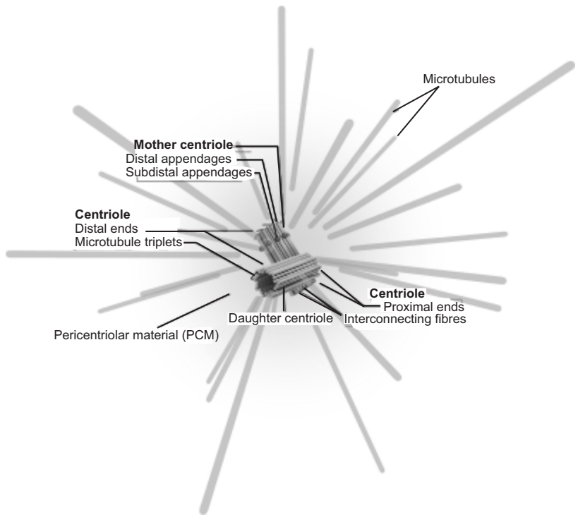
10. Centrosome:
Centrosomes are associated with the nuclear membrane during the prophase of the cell cycle. In mitosis, the nuclear membrane breaks down and the centrosome nucleated microtubules (parts of the cytoskeleton) can interact with the chromosomes to build the mitotic spindle.
11. Chromosomes:
These play a very important role in heredity, mutation and variation. Chromosomes have the capacity for self-reproduction.
Golgi complex is present in all eukaryotic cells except mammalian red blood corpuscles. In-plant cells as well as in invertebrate tissues, it is not present as a single body but there are many such organelles scattered throughout the cytoplasm. Lysosomes are present in animal cells and not in plant cells. Plastids are organelles that are only present in plant cells. They are absent in bacteria and blue-green algae.
Cell Division
Types of Cell Division (Mitosis and Meiosis): Cell division includes mitosis and cytokinesis.
Mitosis
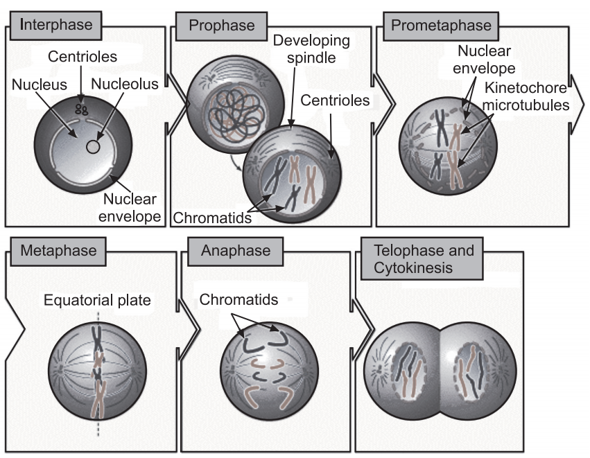
In mitosis, the cell nucleus is replicated and divided into two identical nuclei containing genetically identical material. The four main phases of mitosis are:
1. Prophase:
It is the first step that follows the G2 phase of the interphase.
Characteristics of Prophase:
- Chromosomal material condenses and is seen to be composed of two chromatids and centromere.
- The nuclear envelope breaks up. Spindle fibres begin to form and extends from centrioles. These are made up of microtubules and attached to the centromere of sister chromatids.
- Centrioles slowly migrate to the opposite side of the cell.
- At the end of prophase, cells do not show under microscope, Golgi complex, endoplasmic reticulum, nucleolus and nuclear envelope.
2. Metaphase:
Characteristics of Metaphase:
- In this phase, chromosomes move and lined up along the equatorial plate or metaphase. (equatorial plate is an imaginary line in the centre of the cell.)
- In this phase, chromosomes move with the help of spindle fibres and centrioles. The spindle fibres are attached to kinetochores (disc-shaped structures at the centromere and serve as the site of attachment of spindle fibres) of chromosomes.
3. Anaphase:
Each chromosome, arranged at metaphase, is split and pulled apart by spindle fibres, causing sister chromatids to separate and create two daughter chromosomes. The daughter chromosome further migrates towards opposite poles. This ensures that daughter cells will have identical sets of chromosome and is also identical to the parent cell.
4. Telophase:
New nuclei begin to form around a new set of chromosomes. Spindle fibre disappears at the end of this phase and chromosomes cluster at opposite poles and back into their loose form. The nuclear envelope assembles around the chromosome and nucleolus, Golgi complex and ER reform.
Significance of Mitosis:
Mitosis results in the production of diploid daughter cells with the identical genome (some lower plants also divide by mitosis). The growth of multicellular organisms is due to mitosis. It restores the nucleo-cytoplasmic ratio and is also significant in cell repair. Mitotic division in meristematic tissues, at apical and lateral cambium, results in the continuous growth of plants throughout their life.
Cytokinesis: At the same time cytokinesis begins to form two new cells. In cytokinesis, cell cytoplasm divides into two, making two new daughter cells. In-plant cells, a cell plate begins to form along the centre of the cell and then fuses with the cell membrane.
At the time of cytoplasmic division organelles like mitochondria, plastids get distributed between two daughter cells.
Meiosis
Meiosis is the formation of gametes (production of haploid cells) from specialized diploid cells. This is performed by reproductive cells only. Meiosis involves two cycles of nuclear and cell division (Meiosis I and Meiosis II) and only a single cycle of DNA replication.
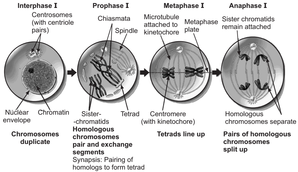
Meiosis I:
It is a four-step process:
1. Prophase I: It is a longer and more complex process as compared to the prophase of mitosis. It is further divided into more steps where chromosomes thicken and are visible under a microscope. Then chromosomes start pairing (synapsis) and are known as homologous chromosomes. These homologous chromosomes move towards the equatorial plate. Thus, four sister chromatids move together and are called “tetrad”. During this time, the nuclear envelope disappears and spindle fibres begin to form (crossing over phase). Crossing over leads to the recombination of genetic material on two chromosomes. At the end of prophase I, tetrad separates except the site of cross over. These X-shaped structures are called “chiasmata”.
2. Metaphase I: During this stage, homologous pairs are lined up along the equatorial plate.
3. Anaphase I: The homologous chromosomes separate, while sister chromatids remain associated at their centromeres (each chromosome has two sister chromatids).
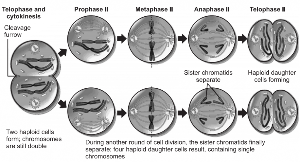
4. Telophase I: The nuclear membrane and nucleolus reappear and then this follows cytokinesis resulting in two new cells each with a haploid number of chromosomes and are in the form of sister chromatids.
This follows Meiosis II.
Meiosis II:
Meiosis II resembles normal mitosis. It is initiated immediately after cytokinesis through four steps namely – prophase II, metaphase II, anaphase II and telophase II.
Prophase II: Nuclear membrane disappears at the end of prophase II.
Metaphase II: During this stage, chromosomes get aligned at the equator and chromosomes move with the help of spindle fibres.
Anaphase II: Each chromosome split and get pulled by spindle fibres allowing them to move towards opposite poles of the cell.
Telophase II: During this phase, new nuclei begin to form and chromosomes once again get enclosed by the nuclear envelope. This follows final cytokinesis resulting in the formation of four haploid daughter cells.
Significance of Meiosis:
Conservation of the specific chromosome number of each species is achieved across generations in sexually reproducing organisms. It also increases genetic variability from one generation to the next. Such variations are important for the evolution process.
Make sure you also check our other amazing article on : Plant Respiration