
Types of Electron Microscopes: An electron microscope (EM) is an instrument, which utilizes a short wavelength of electrons as a source of illumination for observing objects at a greater magnification. The major significance of an electron microscope is that it has the highest resolution and magnification. German engineers Max Knoll and Ernest Ruska 1931, developed the first electron microscope.
The electron microscope works on a principle similar to that of a light microscope. An electromagnetic field and a beam of electrons act in a way similar to the action of a glass lens and a beam of light. The better resolution of electron microscopes is due to the shorter wavelengths of electrons. The wavelengths of electrons are about 100,000 times smaller than the wavelengths of visible light. Electron microscopes are used to examine structures too small to be resolved with light microscopes. It is possible to resolve objects as small as 10 Ao by electron microscopy.
Types of Electron Microscopes:
- Transmission electron microscope (TEM).
- Scanning electron microscope (SEM).
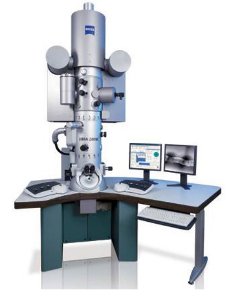
Transmission Electron Microscope
In a transmission electron microscope (Fig.1), a beam of electrons is projected from an electron gun and is passed through a series of electromagnetic lenses (Fig.2). The electron beam is produced by an electron gun, commonly fitted with a tungsten filament cathode as the electron source. The electrons accelerated by the high voltage energy are forced through a collimating aperture which renders the rays in parallel lines and fashions them into a beam. The beam is focused on a small area of the specimen by an electromagnetic condenser lens. The condenser lens is a magnetic coil that corrects the aberrations (bending) in the beam. Electrons get scattered, transmitted through the object, and pass through the objective lens which magnifies the image of the object (Fig.3).
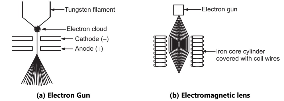
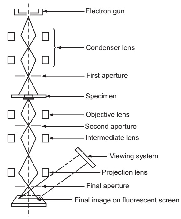
The strength of the magnetic lens depends on the amount of current that is allowed to flow through it. The electron beam that has been partially transmitted through the very thin specimen carries information about the structure of the specimen. The entire electron microscope must be in a vacuum or otherwise the electrons get scattered due to collisions with air molecules and fail to get focused. The specimen must be absolutely dry. Specimen supporting grid is usually a collodion film or Fonnvar or polymerized plastic plus a screen grid of copper. The electron beam that passes through an object is scattered depending on the varying refractive index of the specimen. The electron beam from the specimen passes through the second set of magnetic coils (objective lens), which focus the electrons and form an intermediate image. A third set of magnetic lenses (projection lens), further magnifies the image and projects it on the fluorescent screen or photographic film. The electron image is converted into visible form by projecting it on a fluorescence screen.
An electron beam has low penetration power through solid matter. Hence, very thin sections of the specimen can be examined under an electron microscope. The degree of scattering of electrons by the specimen is related to the number and mass of the atoms that lie in the electron path. Since most of the constituent elements in the biological matter are of low mass and the contrast of these materials is weak. The contrast of such materials can be enhanced by staining with salts of heavy metals such as uranium or tungsten. These metals may be fixed on the specimen (positive staining) or used to increase the opacity of the surrounding area (negative staining).
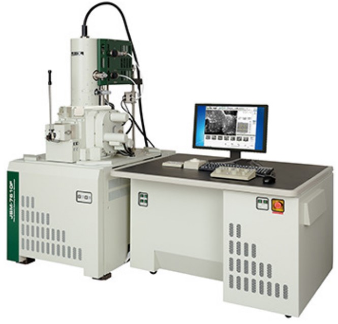
Scanning Electron Microscope
The scanning electron microscope (SEM) was built by Van Ardene in 1938. SEM (Fig.4) is primarily used for visualizing the surface architecture of the specimen rather than the internal details. An SEM provides striking three-dimensional views of specimens. The construction plan and working principle of an SEM are different from that of a TEM.
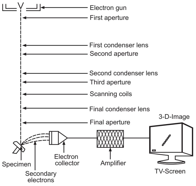
In SEM, an accelerated beam of electrons is produced from the electron gun and is focused on the specimen by the condenser lens. The magnetic lenses of an SEM are responsible to produce an extremely thin beam of electrons called the primary electron beam. These electrons pass through electromagnetic lenses and are directed over the surface of the specimen (Fig.5). The primary electron beam knocks electrons out of the surface of the specimen. This causes the release of secondary electrons from the specimen surface. The intensity of these secondary electrons depends on the shape and the chemical composition of the irradiated object. The secondary electrons are collected by a detector which generates an electron signal. The signals are then scanned in the manner of a television system to produce an image on a Cathode Ray Tube (CRT).
During this process, some of the primary electrons are also reflected and transmitted to the collector but their number is less than the secondary electrons. As a result, the image signal is developed more by the secondary electrons than the primary electrons. The secondary electrons deflected out of the specimen will be a replica of the refractive index of the surface and thus produce an image on the CRT screen revealing all the topographical details. Image contrast is mainly dependent on surface topography which determines the number of secondary electrons reaching the detector. SEM also has a resolution equal to that of TEM. A resolution from 1 to 10 nm is possible with a corresponding magnification from 10,000 to 100,000. This microscope is especially useful in studying the surface structures of intact cells and viruses. In the pharmaceutical field, SEM is very useful in studies associated with the surface characteristics of drug particles and morphological studies of antibiotic-producing microorganisms and their spores.
Limitations of Electron Microscopy:
- The specimen being examined is in a chamber that is under a very high vacuum. Thus, cells cannot be examined in a living state.
- The drying process may change some morphological characteristics.
- Thin sections are required to observe the internal structures of the cell because of the low penetration power of the electron beam.
- The numerical aperture of an electron microscope lens is very small.
Make sure you also check our other amazing Article on : Compound Microscope