The virus is a small nucleoprotein complex and infectious agent replicates only inside the living cells of other organisms such as animals and plants. They are much smaller than bacteria (20-300 nm). They are obligate intracellular parasites of bacteria, protozoa, fungi, algae, plants, and animals. In 1890, D. Ivanovski and M. Beijerinck discovered Tobacco mosaic virus that caused disease in the tobacco plant. Friedrich Loeffler and Paul Frosch have discovered animal viruses that cause foot and mouth disease in cattle. They cannot be grown in a sterile medium but grow in specific host cells. They have only a single type of nucleic acid that serves as genetic material but lack cellular structure and enzymes for metabolite processes. They are highly species-specific.
Properties of Viruses:
- They do not have a cellular organization.
- They contain only one type of nucleic acid either DNA or RNA.
- They are obligate intracellular parasites.
- They lack enzymes for protein synthesis.
- They multiply by the complex process but not by binary fission.
- They are unaffected by antibacterial antibiotics.

Morphology
Table of Contents
Viruses are acellular that means they do not have cells as it has no nucleus, cytoplasm or organelles. They consist of two main parts viz. nucleic acid and capsid. In nucleic acid, DNA or RNA is found at the core of the virus. They need a living host to survive. The smallest infectious virus is 20 nm to 300 nm. The smallest RNA virus is a picornavirus, the smallest DNA virus is parvovirus (20 nm) and the largest virus is poxvirus (300 nm). They may be single or double-stranded. The nucleic acid is linear or circular and either segmented or non-segmented. A capsid is a protein coat that is surrounded by genetic material. A protein coat is made up of repeating sub-units known as capsomeres. Based on the arrangement of the morphological sub-unit, viruses are grouped into three types namely Icosahedral symmetry, Helical symmetry, and Complex symmetry.
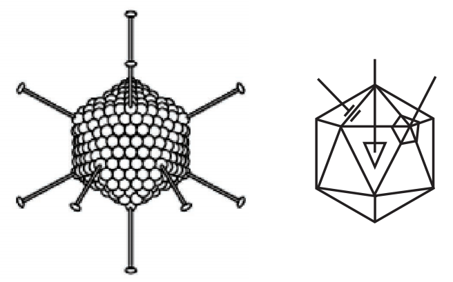
(a) Icosahedral symmetry: Capsomeres are arranged in 20 triangles (Fig.1), Example: Adenovirus.
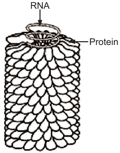
(b) Helical symmetry: They are arranged in a hollow coil that appears rod-shaped (Fig.2), Examples: Influenza virus, Rabies virus, etc. and
(c) Complex symmetry: They have complexity in the structure of viruses and they do not show icosahedral or helical symmetry. Example: Poxviruses.
Some viruses have an outer envelope that is made of lipids and proteins covering the capsid. They are parasites. Most viruses are spherical shaped but some are different types like pox virus is brick-shaped, the bacteriophage is tadpole-shaped, tobacco mosaic virus is rod-shaped. They do not have ribosomes and do not undergo mitosis or binary fission.
Functions of Viral Capsid:
- It protects genetic materials.
- It serves as antigenic determinants.
- It induces antibody production.
- It provides the structural symmetry of the virus.
- It facilitates the assembly and packaging of viral genetic information.
- It serves as a vehicle of transmission from one host to another.
Viral Envelope:
It is a lipoprotein derived from the host cell membrane and virus-specific protein. It is composed of glycoproteins that look like spikes. It confers instability to the virus because of the loss of infectivity due to the disruption of lipid. They are more sensitive to heat, lipid solvents, and detergents.
Functions:
- They help to attach to host cell receptors.
- They are antigenic determinants.
- They stimulate antibody production.
Examples: Herpes virus, HIV, Hepatitis virus, etc.
Virion:
It is an entire virus particle, consisting of an outer protein shell called a capsid and an inner core of nucleic acid. They are present in naked viruses, identical to nucleocapsid whereas in enveloped viruses develops envelop.
Peplomers:
In mature virus particles, the glycoprotein often appears as projecting spikes on the outer surface of the envelope, which is known as peplomers. Example: Influenza virus carries two types of peplomers viz. hemagglutinin and neuraminidase.
Functions:
- It helps for the attachment of the virus to the host cell receptors to initiate the entrance of the virion into the cell.
- It has antigenic properties.
- It has an enzymatic activity like neuraminidase which cleaves neuraminic acid from host cell glycoproteins.
- It attaches to the receptors on the RBC, causes these cells to agglutinate.
Classification of Viruses (Flowchart.1)
Viruses are classified by phenotypic characteristics, such as morphology, nucleic acid type, mode of replication, host organisms, and the type of disease they cause. They are as follows:
1. Classification based on nucleic acid
2. Classification based on structure or symmetry
3. Classification based on replication properties and site of replication
4. Classification based on host range
5. Classification based on the mode of transmission
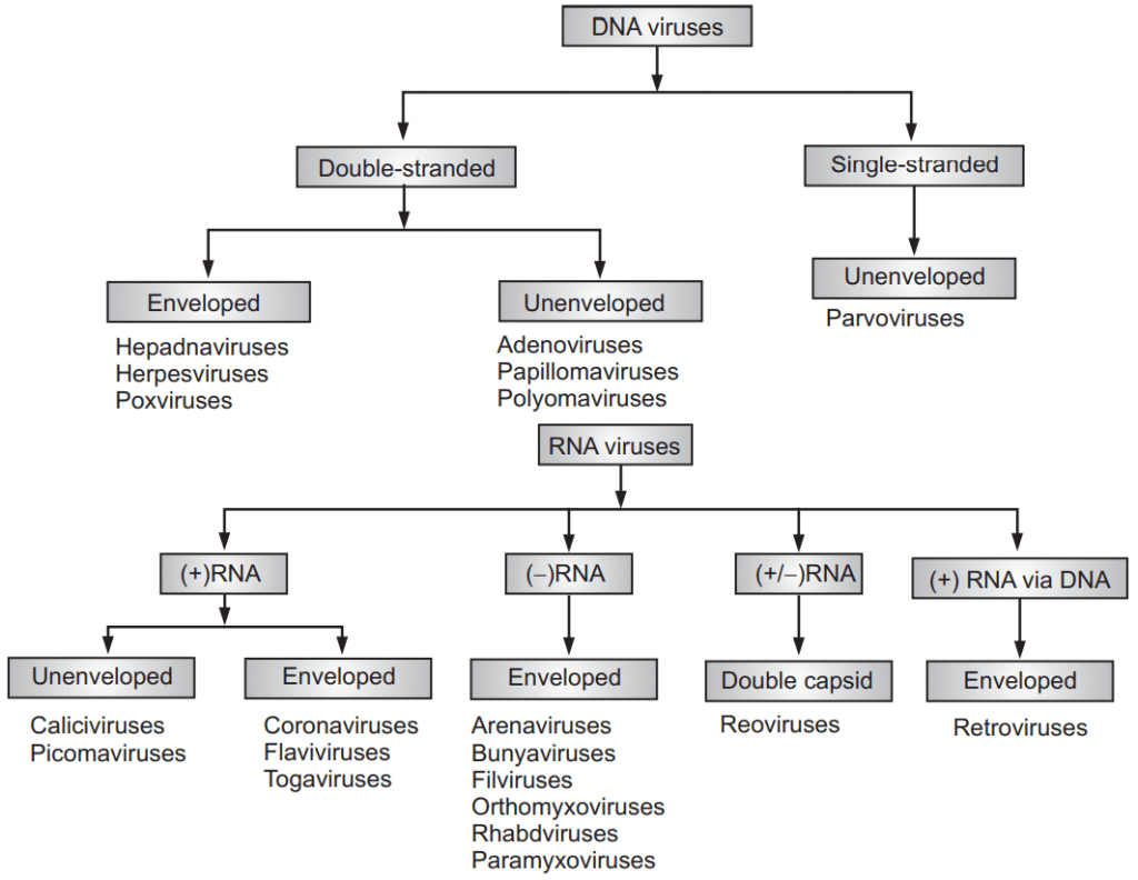
1. Classification based on Nucleic Acid
(a) DNA virus:
The viral genome is DNA.
(i) Double-stranded DNA virus: Examples: Adenovirus, Herpesvirus.
(ii) Single-stranded DNA virus: Example: Parvovirus.
(b) RNA virus:
Genome is RNA.
(i) Double-stranded RNA virus: Example: Reovirus.
(ii) Single-stranded RNA virus: These are further classified into two groups.
- Positive sense RNA (+RNA): Polio virus, Influenza virus.
- Negative sense RNA (−RNA): Rabies virus
2. Classification of Virus based on Structure
(a) Cubical virus:
- They are also known as icosahedral symmetry viruses.
- Examples: Reovirus, Picorna virus.
(b) Spiral virus:
- They are also known as helical symmetry viruses.
- Examples: Paramyxovirus, orthomyxovirus.
(c) Radial symmetry virus:
- They look like spoke or wheel type.
- Example: Bacteriophage
(d) Complex virus:
- They possess a capsid that is neither purely helical nor purely icosahedral.
- Example: Pox virus.
3. Classification of the virus based on replication properties and site of replication
(a) Replication and assembly in the cytoplasm of host:
- Example: All RNA viruses replicate and assemble in the cytoplasm of host cells except the Influenza virus.
(b) Replication in nucleus and assembly in the cytoplasm of host:
- Examples: Influenza virus, Pox virus.
(c) Replication and assembly in the nucleus of host:
- All DNA viruses replicate and assemble in the nucleus of the host cell except the Pox virus.
(d) Virus replication through ds DNA intermediate:
- Examples: All DNA viruses, Retrovirus, and some tumor-causing RNA viruses replicate through ds DNA as intermediates.
(e) Virus replication through ss RNA intermediate:
- Example: All RNA viruses except Reovirus and tumor-causing RNA viruses.
4. Classification of Virus based on Host Range:
(a) Bacteriophage:
- Phages are viruses infecting bacteria. Examples: λ phage, T2, T4, φ174, MV-11.
(b) Plant virus:
- Those viruses infect plants. Examples: TMV, cauliflower mosaic virus.
(c) Animal virus:
- Those viruses that infect animals. Examples: Poliovirus, Retrovirus, Herpes virus, Adenovirus.
(d) Insect virus:
- The virus that infects insects. Examples: Baculovirus, Sacbrood virus, Entomopox virus, Granulosis virus
5. Classification of virus based on the mode of transmission:
(a) Virus transmitted through respiratory route:
- Examples: Swine flu, Rhinovirus.
(b) Virus transmitted through fecal-oral route:
- Examples: Hepatitis A virus, Poliovirus, Rotavirus.
(c) Virus transmitted through sexual contacts:
- Example: Retrovirus.
(d) Virus transmitted through blood transfusion:
- Examples: Hepatitis B virus, HIV
(e) Zoonotic virus:
- Viruses are transmitted through the biting of infected animals. Examples: Rabies virus, Alphavirus, Flavivirus.
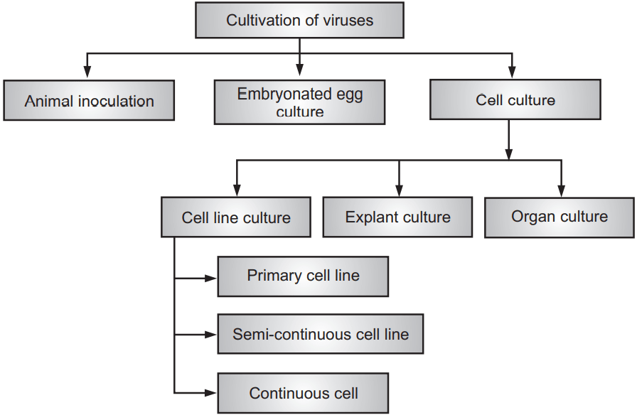
Cultivation of Viruses
Viruses are cultured in embryonated egg, cell line culture, and animal inoculation because they can only replicate inside the host cell, so viruses cannot be cultured in the non-living medium as bacteria and fungi (Bacteria and fungi are living creatures). The main purposes of virus cultivation are identification and isolation of viruses in clinical samples, research on the viral structure, replication, genetics, and their effects on the host cell, and production of viral vaccines. Hence, the cultivation of viruses is essential which is carried out by three methods viz. Animal inoculation, Embryonated egg culture, and Cell culture (Flowchart 2).
1. Animal inoculation:
Animal inoculation is one of the primary methods for isolating certain viruses and studying the pathogenesis of certain viral diseases. This method is carried out for those viruses which are not cultivated in embryonated egg and tissue culture methods. In this method, healthy and disease-free laboratory mice (white mice) are particularly used for virus cultivation. Suckling mice of age less than 48 hours are used for culture of Toga virus and Coxsackie virus. The inoculation of viruses is carried out by the intracerebral and intranasal routes. The inoculation is also carried out by intraperitoneal and subcutaneous routes. Other healthy and disease-free animals such as hamsters, Guinea pigs, rabbits, chimpanzees, and primates are also used as an alternative for virus culture. After inoculation of virus sample into the host cell, the animals are observed for symptoms of disease till death and then the virus is isolated from tissue of an animal. Live inoculation was first used to study of yellow fever virus on human volunteers.
Advantages:
- Diagnosis, Pathogenesis, and clinical symptoms are determined.
- Antibodies productions are identified.
- Mice are the reliable model for virus replication.
- This method helps to study immune responses, epidemiology, and oncogenesis.
Disadvantages:
- The animal experiment is costly and difficult to maintain.
- Animal selection for specific viruses is difficult.
- Some human viruses are not grown in animals.
- Vaccine production is not possible with the mice model.
2. Embryonated egg culture:
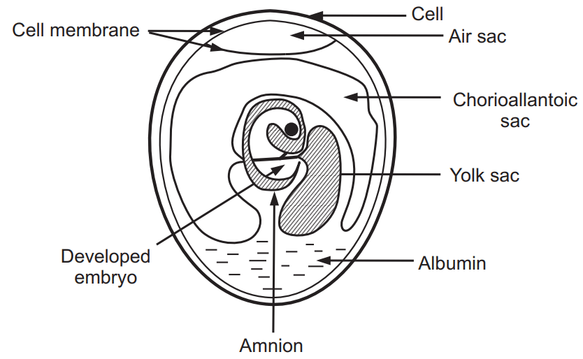
For virus cultivation, an egg embryo of 7-12 days is used. At first, the egg is kept in an incubator for embryo development up to 7-12 days and then the virus sample is inoculated into the egg. For inoculation, eggs are first prepared for cultivation and the shell surface is first disinfected with iodine and penetrated with a small sterile drill. After inoculation, the opening is sealed with paraffin and incubated at 36°C for 2-3 days. The egg is broken after incubation and the virus is isolated from the tissue of the egg. The virus can be cultured in different parts of the embryonated egg, such as chorioallantoic membrane (Poxvirus are cultured), amniotic sac (Influenza virus, Mumps virus, Yellow fever virus, and Rabies virus are cultivated), amniotic cavity (Influenza virus is cultured) or yolk sac (Herpes virus is cultured) depending upon types of virus. In 1931, Scientist Pasture was first used the embryonated hen’s egg for the cultivation of virus (Fig. 3).
Advantages:
- This method is a widely used method for the isolation of the virus.
- It is an ideal substrate for viral growth and replication.
- This method is cost-effective and maintenance is much easier.
- Less labor is needed.
- The embryonated eggs are readily available.
- They are free from contaminating bacteria and many latent viruses.
Disadvantages:
- Each virus has different sites for its growth and replication hence the site of inoculation varies with the viruses.
3. Cell culture (tissue culture) technique:
This technique is the most commonly used technique for the cultivation of viruses. There are three types of cell culture techniques viz. organ culture, explant culture, and cell culture.
(i) Organ culture: This culture method is mainly done for highly specialized parasites of certain organs. Like, tracheal ring culture is done for isolation of coronavirus.
(ii) Explant culture: In this method, a small fragment of tissue is extracted from humans or animals and used for virus culture but this technique is very rarely used.
(iii) Cell line culture: This is the most commonly used technique. Cell line culture is routinely used in the lab for virus culture, isolation and identification. In this method, the growth media is prepared by maintaining a balanced salt concentration, all essential amino acids, glucose, buffering agents, some antibiotics, serum, etc. Thereafter some tissue fragment is obtained which is trypsinized to dissociate cells. These cells are washed and suspended in culture media in Petri plates and incubated for a sufficient time. On incubation, the cell divides and spreads out on the glass surface to form a confluent monolayer of cells that are used for virus culture.
Based on origin, chromosomal characteristics, and the number of generations through which cell culture can be maintained, cell line culture is classified into three types viz. Primary cell line, Semi-continuous cell line, and Continuous cell line.
I. Primary cell line:
These are normal cells, obtained from the fresh organ of animals or humans and cultured. These primary cell line cultures are used for the isolation of viruses and the preparation of vaccines. These cells are capable of limited growth for a limited generation. They cannot be maintained in a serial sub-culture. Examples: Monkey kidney cell line, Human amnion cell line, etc.
II. Semi-continuous cell line:
These cells are fibroblastic or also known as diploid cells because they contain the same number of chromosomes as the parent cell. These diploid cells are sub-cultured for a limited generation. They are susceptible to a wide range of Human virus cultures and are also used for vaccine production. Examples: Rhesus embryo cell, human embryonic lung strain, etc.
III. Continuous cell line:
These are cells of a single type capable of infinite growth in-vitro. These are usually cancer cells derived from cancerous tissue. These cells grow faster and they are haploid cells. These cells are maintained by serial sub-culture or by deep freezing at −7°C so that these cells are reused when necessary. This cell line is used for virus culture but not used for vaccine preparation. Examples: HeLa cell is obtained from cervical cancer, HEP-2 (Human Epithelioma of larynx cell line), Vero (Vervet monkey) kidney cell lines, BHK-21 (Baby Hamster Kidney cell line).
Advantage of Cell Culture:
- The method is easy, broad-spectrum, cheaper, and good sensitivity.
Disadvantages of Cell Culture:
- The process requires a skilled person.
- Tissue or serum is sent to central laboratories for analysis to identify the virus.
Cultivation of Bacteriophages
Bacteriophages are cultivated in either broth or agar cultures of young, actively growing bacterial cells.
Cultivation of Plant Viruses:
Plant viruses are cultivated by plant tissue cultures, cultures of separated cells, or cultures of protoplasts, etc., and viruses are grown in whole plants. Leaves are mechanically inoculated by rubbing with a mixture of viruses and an abrasive and that time cell wall is broken and the viruses directly contact the plasma membrane and infect the exposed host cells. A localized necrotic lesion often develops due to the rapid death of cells in the infected area.
Reproduction of Viruses
Viruses do not reproduce. The living cell in which the virus reproduces is called a host cell. They use these host cells to replicate themselves by creating an exact copy of the virus. Two main types of reproductive cycle are observed in viruses namely Lytic Cycle and Lysogenic Cycle.
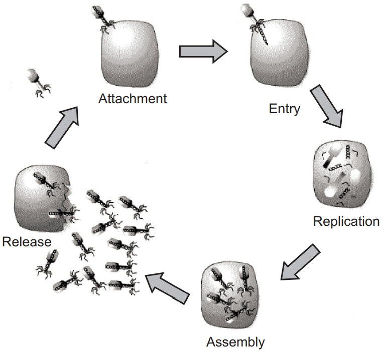
Lytic Cycle (Fig. 4): This cycle is completed with several steps which are as follows:
1. Attachment: In this first step, Virus is attached to the host cell.
2. Entry: Then genetic material is injected into the host cell.
3. Replication: The virus takes over the cell’s metabolism, causing the creation of new proteins and nucleic acids by the host cell’s organelles.
4. Assembly: Then proteins and nucleic acids are assembled into new viruses.
5. Release: Finally virus enzymes cause the cell to burst and viruses are released from the host cell. These new viruses further infect other cells and this process is continuing.
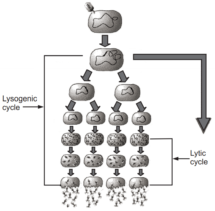
Lysogenic Cycle (Fig. 5): This cycle is completed with several steps which are as follows:
1. Attachment: In the first step, a virus is attached to the host cell.
2. Entry: Then genetic material is injected into the host cell.
3. Integration: Viral DNA integrates into the host cell’s genome.
4. Replication (lysogenic cycle): When the host cell replicates, viral DNA is copied along with host cell DNA. Each new daughter cell is infected with the virus.
5. Induction: When the infected cells are exposed to certain environmental conditions, viral DNA is activated and enters the lytic cycle.
6. Replication (lytic cycle): The virus takes over the cell’s metabolism, causing the creation of new proteins and nucleic acids by the host cell’s organelles.
7. Assembly: Proteins and nucleic acids are assembled into new viruses.
8. Release: Finally, virus enzymes cause the cell to burst, and viruses are released from the host cell. These new viruses can infect other cells.
Make sure you also check our other amazing Article on : Fungi