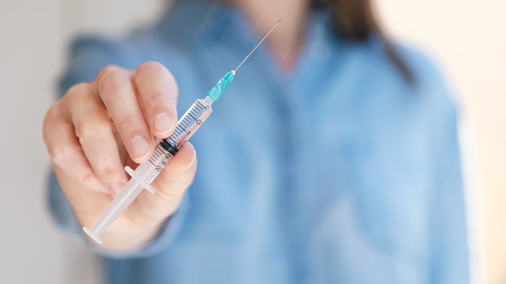
Parenteral Dosage Form: Parenteral refers injectable route of administration. It is derived from the Greek words Para (Outside) and enteron (Intestine). So it is a route of administration other than the oral route. This route of administration bypasses the alimentary canal Pyrogens, fever-producing substances are primarily lipid polysaccharide products of metabolism of microorganisms; they may be soluble, insoluble, or colloid.
Parenteral Dosage Form:
Table of Contents
1. Parenteral preparations must be sterile – free of microorganisms.
2. To ensure sterility, parenteral are prepared using
- aseptic techniques
- special clothing (gowns, masks, hair net, gloves)
- laminar flow hoods placed in special rooms.
Advantages of the Parenteral Route:
1. The IV route is the fastest method for delivering systemic drugs
- preferred administration in an emergency.
2. It can provide fluids, electrolytes, and nutrition
- patients who cannot take food or have serious problems with the GI tract.
3. It provides a higher concentration of drug to the bloodstream or tissues
- advantageous in serious bacterial infection.
4. IV infusion provides a continuous amount of needed medication without fluctuation in blood levels of other routes.
5. Infusion rate can be adjusted
- to provide more or less medication as the situation dictates, drug action can be prolonged by modifying the formulation.
Disadvantages of the Parenteral Route:
1. Traumatic injury from the insertion of the needle.
2. Potential for introducing:
- Toxic agents
- Microbes
- Pyrogens
3. Impossible to retrieve if adverse reaction occurs.
- injected directly into the body.
4. Correct syringe, needle, and technique must be used.
5. Rotation of injection sites with long-term use
- prevents scarring and other skin changes.
- can influence drug absorption.
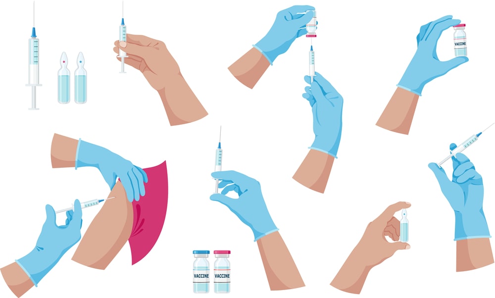
Routes of Administration of Parenteral Products
Various types of the route of administration of Parenteral products are:
Intradermal Injection:
- Subcutaneous (Hypodermis) injection
- Intramuscular injection
- Intravenous injection
- Intra-arterial injection
- Intracardiac injection
- Intrathecal injection
- Intracisternal injection
- Peridural injection
- Intra-articular injection
- Intracerebral injection
Subcutaneous Injections:
- Given at a 45-degree angle 25- or 26-gauge needle, 3/8 to 5/8 inch length.
- No more than 1.5 ml should be injected into the site to avoid pressure on sensory nerves causing pain and discomfort.
- Administer medications below the skin into the subcutaneous fat outside of the upper arm top of the thigh lower portion of each side of the abdomen, not into grossly adipose, hardened, inflamed, or swollen tissue.
- Often have a longer onset of action and a longer duration of action compared with IM or IV injection.
Intramuscular Injections:
- Care must be taken with deep IM injections to avoid hitting a vein, artery, or nerve.
- In adults, IM injections are given into the upper, outer portion of the gluteus maximus large muscle on either side of the buttocks.
- For children and some adults, IM injections are given into the deltoid muscles of the shoulders.
- The typical needle is a 22-25 gauge ½- to the 1-inch needle.
- IM injections are administered at a 90o angle volume limited to less than 3 ml.
Intravenous Injections or Infusions:
- Fast-acting route because the drug goes directly into the bloodstream often used in the emergency department and critical care areas.
- Commonly used for fluid and electrolyte replacement to provide necessary nutrition to the patient who is critically ill.
- Intravenous (IV) injections are administered at a 15o to 20o angle.
Intra Arterial Injections:
- These injections are given directly into the artery.
Intracardiac injections:
- These are given into the heart muscle or ventricle at the time of emergency only.
Intrathecal Injections:
- These are given into the subarachnoid space the surrounds the spinal cord. This route is used for spinal anesthesia.
Official types of injections:
1. Injection: Liquid preparations that are drug substances or drug solutions thereof e.g. insulin injection USP.
2. For injection: Dry solid that, upon addition of suitable vehicles yield solutions confirming in all respect to the requirements to the injection. e.g. Cefuroxime injection USP.
3. Injectable emulsions: Liquid preparation of drug substance dispersed in a suitable emulsion medium. e.g. Propofol USP.
4. Injectable suspension: Liquid preparation of solid suspended in a suitable medium. e.g. Methyl Prednisolone Acetate Suspension USP.
5. For injectable suspension: Dry solid that upon addition of suitable vehicle yields preparation confirming in all respect to the requirements for Injectable suspension. e.g. Imipenem and Cilastatin Injectable suspension.
General Requirements of Parenteral Preparations:
- Stability
- Sterility
- Free from pyrogens
- Free from foreign particles
- Isotonicity
- Specific gravity
- Chemical purity
Formulation of Parenteral Products:
In the preparation of parenteral products, the following substances are added to make a stable preparation:
1. The active drug
2. Vehicles:
- Aqueous vehicle (e.g. water for injection, water for injection free from CO2)
- Non-aqueous vehicle (e.g. Ethyl alcohol, propylene glycol, almond oil)
3. Adjuvant:
- Solubilizing agents (e.g. Tweens and polysorbates)
- Stabilizers and antioxidants (e.g. thiourea, ascorbic acid, tocopherol)
- Buffering agents (e.g. citric acid, sodium citrate)
- Antibacterial agents (e.g. benzyl alcohol, metacresol, phenol)
- Chelating agents (e.g. EDTA)
- Suspending, emulsifying, and wetting agents (e.g. MC, CMC)
- Tonicity factor (e.g. sodium chloride, dextrose)
Processing of Parenteral Preparation
Following steps are involved in the processing of parenteral preparation:
1. Cleaning of containers, closures, and equipments.
2. Collection of materials.
3. Preparation of parenteral products.
4. Filtration.
5. Filling the preparation in the final container.
6. Sealing the container.
7. Sterilization
8. Evaluation of the parenteral preparation.
9. Labelling and packaging.
1. Cleaning of containers, closures, and equipments: Thoroughly cleaned with detergents with tap water distilled water finally rinsed with water for injection. Rubber closures are washed with 0.5% sodium pyrophosphate in water.
2. Collection of materials: All raw material of preparation should be collected from the warehouse after being accurately weighed. Water for injection should be Pyrogens-free.
3. Preparation of parenteral products: The Parenteral preparation must be prepared in aseptic conditions. The ingredients are accurately weighed separately and dissolved in the vehicle as per the method of preparation to be followed.
4. Filtration: The Parenteral preparation must be filtered by bacteria proof filter such as a filter candle, membrane filter.
5. Filling the preparation in the final container: The filling operation is carried out under strict aseptic precautions.
6. Sealing the container: Sealing should be done immediately after filling in an aseptic environment.
7. Sterilization: For thermostable substances, the Parenteral products are sterilized by autoclaving method at different temperatures and pressure. E.g. 10 lb pressure (115.5o C, or 240o F) for 30 minutes, 15 lb pressure (121.5o C, or 250o F ) for 20 minutes, 20 lb pressure (126.5o C, or 260o F) for 15 minutes. Heat sensitive or moisture-sensitive materials are sterilized by exposure to ethylene oxide or propylene oxide gas.
8. Evaluation of the Parenteral preparation: The following tests are performed to maintain quality control:
- Sterility test
- Clarity test
- Leakage test
- Pyrogen test
- Assay
9. Labelling & packaging
Evaluation of Parenteral Products
- Sterility testing
- Particulate matter monitoring
- Faculty seal packaging or leakage test
- Pyrogens testing
- LAL test
- Assay or drug content uniformity.
Sterility testing:
Definition: It is a procedure carried out to detect and confirm the absence of any viable form of microbes in or on pharmacopeia preparation or product.
Principle: Sterility testing only shows that organisms capable of growing in selected conditions are absent from the fraction of batch that has been tested. If the microorganism is present in the product can be indicated by turbidity in the clear medium.
Objectives of Sterility Testing
- For validation of sterilization process.
- To check the presence of microorganisms in preparation that is sterile.
- To prevent the issue of contaminated products in the market.
Steps involved in Sterility Testing
- Sampling
- Selection of the quantity of the product to be used
- Methods of sterility testing:
(i) Method 1: Membrane filtration method
(ii) Method 2: Direct inoculation method
- Observation and interpretation must be carried out under aseptic conditions.
1. Sampling: The sample must be representative of the whole bulk material and a lot of final containers.
It is mainly followed by two rules:
- A fixed percentage of the final container is selected.
- A fixed number of containers are taken independent of the lot or batch size.
2. Selection of the quantity of the product to be used: Selection of the quantity of the product to be used for sterility testing depends mainly on the volume or weight in the container.
3. Methods of sterility testing :
(i) Membrane filtration method (Method 1):
The membrane filtration method is appropriate for :
- Filterable aqueous preparations
- Alcoholic preparations
- Oily preparations
- Preparations miscible with or soluble in aqueous or oily (solvents with no antimicrobial effect.
All steps of this procedure are performed aseptically in a Class 100 Laminar Flow Hood.
Membrane filter 0.45 µ porosity, Filter the test solution. After filtration removes the filter. Cut the filter into two halves. The first half (for Bacteria) and the Second half (for fungi), Transfer in 100 ml culture media (Fluid Thioglycollate medium). Incubate at 30-35o C for not less than 7 days. Transfer in 100 ml culture media (Soyabeans-Casein Digest medium). Again incubate at 20-25o C for not less than 7 days. Observe the growth in the media.
(ii) Direct inoculation method (Method 2):
- Suitable for samples with small volumes.
- The volume of the product is not more than 10% of the volume of the medium.
- A suitable method for aqueous solution, oily liquids, ointments, and creams.
- The suitable quantity of the preparation to be examined is transferred directly into the appropriate culture medium and incubate for not less than 14 days.

Observation and results:
Culture media is examined during and after the end of incubation.
The following observations are possible:
- No evidence of growth passes the test for sterility.
- There is evidence of growth re-testing is performed the same number of samples, volume, and media as in the original test. No evidence of growth passes the test for sterility.
- There is evidence of growth isolate and identify the organism. Re-testing is performed with twice the number of the sample if No evidence of growth passes the test for sterility. There is evidence of growth Fail the test for sterility.
Make sure you also check our other amazing Article on : Validation