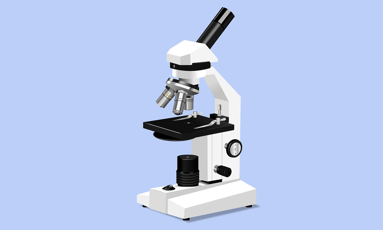
The compound microscope essentially consists of three major systems:
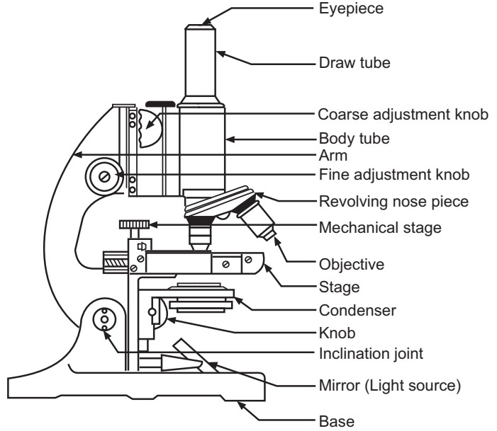
- Support system: It comprises a base, stage, and body tube. (Fig.1)
- Illumination system: It comprises a light source or mirror, an iris diaphragm, and a condenser. The light source may be a plain or concave mirror or electrically illuminated by a tungsten filament lamp or a halogen lamp. A mirror and an electric light source are generally interchangeable.
- Magnification system: It consists of two sets of lenses. The lens system nearest to the specimen is called the objective. The second lens system is called the eyepiece or ocular. The image seen by the eye has a magnification equal to the product of the magnification of the two lens systems.
Important Lenses of Compound Microscope
(A) Oculars:
The functions of the ocular or eyepiece are as follows:
- It magnifies the real image of the object as formed by the objective.
- It corrects some of the defects of the objective.
- It is used for the observation of images.
Different types of eyepieces are used depending upon the kind of objective located on the microscope. The most commonly used oculars are Huygenian, Ramsden, and Compensating.
(a) Huygenian oculars are constructed with the plain surface of two lenses facing upwards and the diaphragm is situated between them at the focus of the upper lens. The lower or field lens collects the rays from as wide a field of view of the image as possible and focusses them at or near the plane of the diaphragm. Huygenian eye-pieces are also called ‘negative oculars’ because the focus occurs within the eye-piece.
(b) Ramsden or positive oculars are constructed with the convex surface of both lenses facing inwards. In this type of ocular, the diaphragm is placed externally below the lower lens. Hence, they are used for micrometry and give more accurate results than Huygenian oculars.
(c) Compensating oculars or positive oculars usually contain a triplet system as the lower lens component. Aberrations are carefully corrected by the makers and they are designed specifically for use with particular objectives.
(B) Objectives:
The objectives are the most important lenses of a microscope because their properties make the final image. The functions of objective lenses are as follows:
- Magnify the real image of the object.
- To unite the light at a point in the image.
- To gather the light rays coming from any point of the object.
There are three major types of objectives mainly used in microscopy such as achromatic, fluorite, and apochromatic. The achromatic objectives are the simplest in construction and the least expensive.
These objectives are used for all the microscopical work of clinical microbiology and most research purposes. Correction of color and spherical aberrations is quite easy in low-power objectives as compared to high-power objectives. Aberrations (rays going away from the right path) are largely eliminated by the use of fluorite and apochromatic objectives. Apochromatic objectives represent the highest degree of optical perfection. These objectives are very costly hence, they are only used for critical research work and photomicrography. Apochromatic objectives are always used with ‘compensating’ eyepieces and a properly centered condenser.
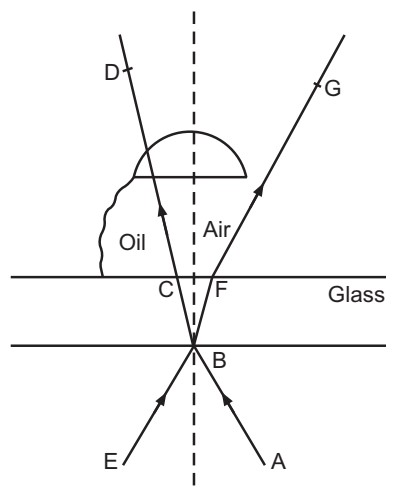
Oil-immersion objectives are most frequently used in microbiology because of their greater magnification and resolution. Most microbial cells are observed with achromatic or apochromatic objectives designed for use with oil immersion. These objectives increase the angle of the cone of rays from the object that enters the objective. With a ‘dry’ objective, the air is present between the object on the surface of the glass slide and the objective lens.
The refractive index of air (n = 1) is lower than that of glass (n = 1.55) and as light rays pass from the glass slide into the air, they are bent or refracted (Fig. 5.6, EBFG) so that they do not pass into the objective lens. This would cause a loss of light which would reduce the numerical aperture and diminish the resolving power of the objective lens. Loss of refracted light can be compensated by using cedar wood oil (n = 1.5), which has the same refractive index as that of glass, between the slide and the objective lens (Fig.2, ABCD). In this way, decreased light refraction occurs and more light rays enter directly into the objective lens producing a vivid image with high resolution.
(C) Condensers:
In microbiology, two methods are commonly used for illuminating the object under the microscope.
1. Illumination by transmitted light: A condenser may be defined as a series of lenses for illuminating transmitted light, an object to be studied on the stage of the microscope. It is located under the stage of the microscope between the mirror and the object. It is also called a substage condenser. A condenser is necessary for the examination of an object with an oil immersion objective to obtain adequate illumination. A condenser is also preferable when working with high-power dry objectives. A good condenser sends light through the object under an angle sufficiently large to fill the aperture of the back lens of
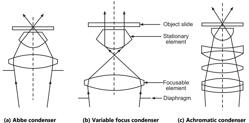
the objective. Therefore, it must be properly positioned during the microscopy. Generally, condensers are also incorporated with an iris diaphragm and a filter holder. An iris diaphragm is used to control light intensity. The Abbe condenser, variable focus condenser, and achromatic condensers are commonly used for bright field illumination.
The Abbe condenser (Fig.3 a) utilizes only two lenses. It is extensively used for general microscopy because of its simplicity and good light-gathering ability. It is not corrected for spherical aberration (which fails to bring the whole microscopic field into simultaneous focus) and chromatic aberration (which produces colored fringes around objects in the field).
The variable-focus condenser (Fig.3 b) is a two-lens system in which the upper lens element is fixed and the lower element is flexible. It is possible to fill the field of low-power objectives without the necessity of removing the top element. This condenser is similar to the Abbe condenser when the lower lens is raised to its top position.
The achromatic condenser (Fig.3 c) is corrected for both chromatic and spherical aberrations. Hence, it is mainly used for research microscopy and for color photomicrography where the highest degree of perfection in the image is desired.
2. Dark field illumination or Dark field microscopy: The microscope which forms a bright image against a dark background is called dark field microscopy. Many transparent and semi-transparent objects are not easily visible in a bright field. Visibility is dependent upon the contrast between the object and its background and can be improved by using a dark background.
The cone of light normally illuminating an object does not enter the objective, but light scattered or reflected by the specimen is seen by the objective. If a dark-field stop of
a suitable size is selected, all the direct rays from the condenser can be made to pass outside the objective. Any object within this beam of light will reflect some light into the objective and be visible.
There are three requisites for adapting an ordinary microscope for dark background illumination.
- A dark background condenser, which focuses only oblique rays of light on the specimen,
- A suitable high-intensity lamp, and
- A funnel stop reduces the numerical aperture of the objective to less than 1.0. The Abbe condenser, paraboloid condenser, and cardioid condenser are commonly used for dark field illumination.
The Abbe condenser (Fig.4 a) is more commonly employed than the other condensers because it is suitable for objects that do not require the highest magnification to make them visible. It may be employed either by inserting a dark-field stop below the condenser or substituting the top part of the condenser with a dark-field element.
The paraboloid condenser is designed to be used with oil-immersion objectives and an intense source of light. The specimen or object must be mounted in a liquid or cement and protected with a cover slip. The numerical aperture of the objective must not be greater than that of the condenser.
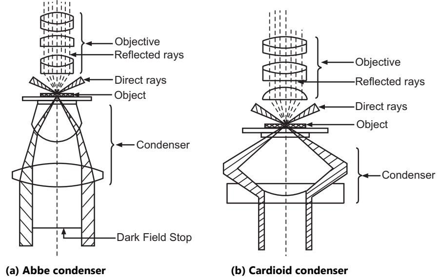
The cardioid condenser (Fig.4 b) is best employed with a strong arc lamp. Ordinary glass slides and cover slips are not used because of the high concentration of light. It is better to employ fused quartz object slides and fused quartz cover slips. The cardioid condenser is specially designed for the examination of colloidal solutions and suspensions.
Dark-field microscopy is very useful for the examination of unstained micro-organisms suspended in fluid i.e. by wet mount techniques and hanging drop preparations. A dark-field examination is useful in the detection of Treponema palladium in the early diagnosis of syphilis.
Make sure you also check our other amazing Article on : Pure Culture Techniques