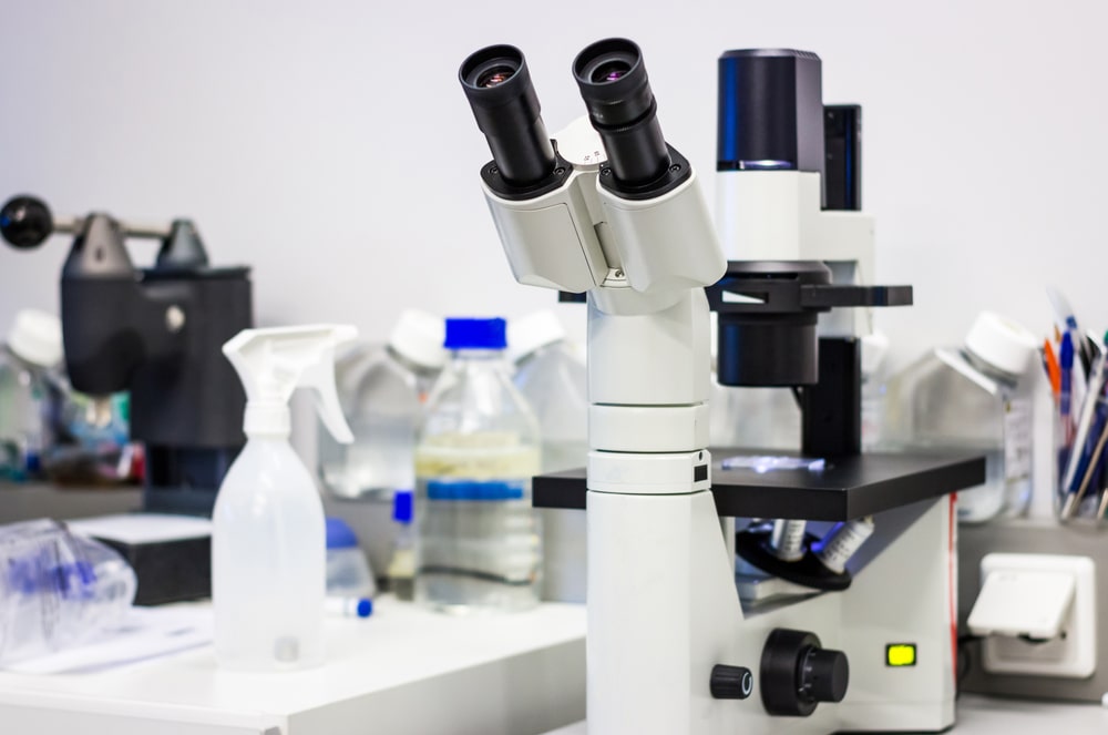
Phase Contrast Microscopy: The phase contrast principle was discovered by Fritz Zernike for which he was awarded the Nobel Prize in Physics in 1953.
The basic construction of a phase contrast microscope is like a bright field microscope except for a special type of condenser and a phase plate. The condenser has a special diaphragm consisting of an annual stop. The annular stop allows only a hollow cone of light rays to pass through the condenser. The phase plate is a special optical disc located in the rear focal plane of the objective. It has a special ring coated with a material that can either advance or retard the direct rays. The rays that pass through the object in a straight line are called direct (undiffracted) rays and are unaltered in amplitude and phase. The rays that are bend and slowed down due to differences in intensity of the medium are called diffracted rays.
The phase of light rays is altered when they pass through a specimen to be observed under a light microscope. This change of phase is a manifestation of the depth and density of the cell and its internal parts. Since there is very little difference or contrast in the refractive indices or density of the specimen, its internal structures, and the medium, it is not made visible by bright-field microscopy. In phase contrast microscopy, the small phase differences are intensified and translated into differences in light intensity with the help of special optical devices. A diagram illustrating an optical system and light transmission through a phase contrast microscope is shown in Figure 1.
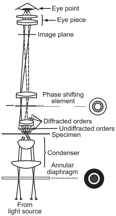
An annular aperture in the diaphragm placed in the focal plane of the sub-stage condenser controls the illumination of the object. The aperture is imaged by the condenser and the objective at the rear focal plane of the objective. In this plane, a phase-shifting disk or phase plate is placed. Undiffracted light rays are transmitted through the object and pass through the altering ring on the phase plate. At this point, they acquire a one-quarter wavelength advance over the diffracted light rays by the object. While diffracted (indirect) rays pass through the transparent region of the phase plate and are unchanged by the missing phase ring they are already retarded by one-quarter (1/4) wavelength due to the object. Finally, all rays including undiffracted rays and diffracted rays are brought together by an eyepiece lens. Apparent brightness or darkness in an image is proportional to the square of the amplitude of light waves. The image will be four times brighter or darker as seen in the bright field microscope. Hence, it is possible to visualize microorganisms without staining.
Phase contrast microscopy provides a second method for observing unstained living microorganisms with good contrast and high resolution. It does not show very small objects like in dark field microscopy but it is more useful for the study of the structure and structural changes in larger microorganisms and tissue cells. Phase contrast microscopy is also useful in the examination of growth and cell division in bacteria, flagellar movements, spore and capsule formation, and the cytopathic effect of viruses in tissue culture.