Introduction of Myocardial Infarction:
Table of Contents
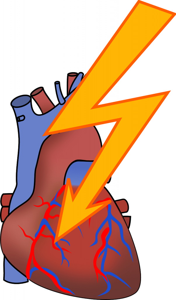
Myocardial Infarction is a condition resulting from decreased blood and oxygen supply to the heart, causing cell death. The major cause is a sudden blockage of coronary arteries.
Coronary arteries are blood vessels that supply the heart muscle with blood and oxygen. Blockage of a coronary artery deprives the heart muscle of blood and oxygen, causing injury to the heart muscle causing chest pain and chest pressure sensation. If blood flow is not restored to the heart muscle within 20 to 40 minutes, irreversible death of the heart muscle will begin to occur. Muscle continues to die for six to eight hours at which time the heart attack usually is “complete.” The dead heart muscle is eventually replaced by scar tissue.
Etiology of Myocardial Infarction:
Myocardial infarction (MI) is the irreversible death (necrosis) of the heart muscle which usually results from an imbalance in oxygen supply and demand, which is most often caused by plaque rupture with thrombus formation in an epicardial coronary artery, resulting in an acute reduction of blood supply to a portion of the myocardium. Acute MI is the single most common cause of death in industrialized nations. Among fatal cases, nearly half of the patients die before reaching the hospital.
Major risk factors include previous cardiovascular disease (such as angina, a previous heart attack, or stroke), older age (especially men over 40 and women over 50), tobacco smoking, high blood levels of certain lipids (triglycerides, low-density lipoprotein, or “bad cholesterol”) and low levels of high-density lipoprotein (HDL, “good cholesterol”), diabetes, high blood pressure, obesity, chronic renal failure, heart failure, excessive alcohol consumption, the abuse of certain drugs (such as cocaine and methamphetamine), and chronic high-stress level.
Pathogenesis of Myocardial Infarction:
1. Myocardial Ischaemia:
Myocardial ischemia is brought about by one or more of the following mechanisms:
- Diminished coronary blood flows e.g. in coronary artery disease shock.
- Increased myocardial demand e.g. In exercise, emotions.
- Hypertrophy of the heart without a simultaneous increase of coronary blood flow e.g. in hypertension, valvular heart disease.
2. Role of platelets:
Rupture of an atherosclerotic plaque exposes the subendothelial collagen to platelets which undergo aggregation, activation, and release reaction. These events contribute to the build-up of the platelet mass that may give rise to emboli or initiate thrombosis.
3. Acute plaque rupture:
In general, slowly developing coronary ischaemia from stenosis coronary atherosclerosis of high grade may not cause acute MI but continue to produce episodes of angina pectoris. But acute complications in coronary atherosclerotic plaques in the form of superimposed coronary thrombosis due to plaque rupture and plaque hemorrhage is frequently encountered in cases of acute MI:
- Superimposed coronary thrombosis due to disruption of plaque is seen in about half the cases of acute MI. Infusion of intracoronary fibrinolysis in the first half an hour of development of acute MI in such cases restores blood flow. The development of acute MI in such cases restores blood flow in the blocked vessel in the majority of cases.
- Intramural hemorrhage is found in about one-third of cases of acute MI.
4. Non-atherosclerotic causes:
About 10% of cases of acute MI are caused by non-atherosclerotic factors such as coronary vasospasm, arteritis, coronary l stenosis embolism, thrombotic diseases, trauma, and outside compression as already described.
The location of an MI is determined by the site of the occlusion and by the anatomy of the coronary circulation. Occlusion of the left anterior descending coronary artery typically causes an infarct in the anterior and apical areas of the left ventricle and adjacent interventricular septum. Occlusion of the right coronary artery is responsible for most infarcts involving the posterior and basal portions of the left ventricle. The underlying anatomy of the coronary circulation also has a significant influence on the location of the infarct. For example, occlusion of the right coronary artery would have different consequences in an individual whose posterior ventricle was supplied by branches of the right coronary than in a person whose posterior wall was supplied by branches of the circumflex coronary artery.
Symptoms of Myocardial Infarction:
Patients with typical MI may have the following symptoms in the days or even weeks preceding the event: Fatigue, Chest discomfort, Malaise.
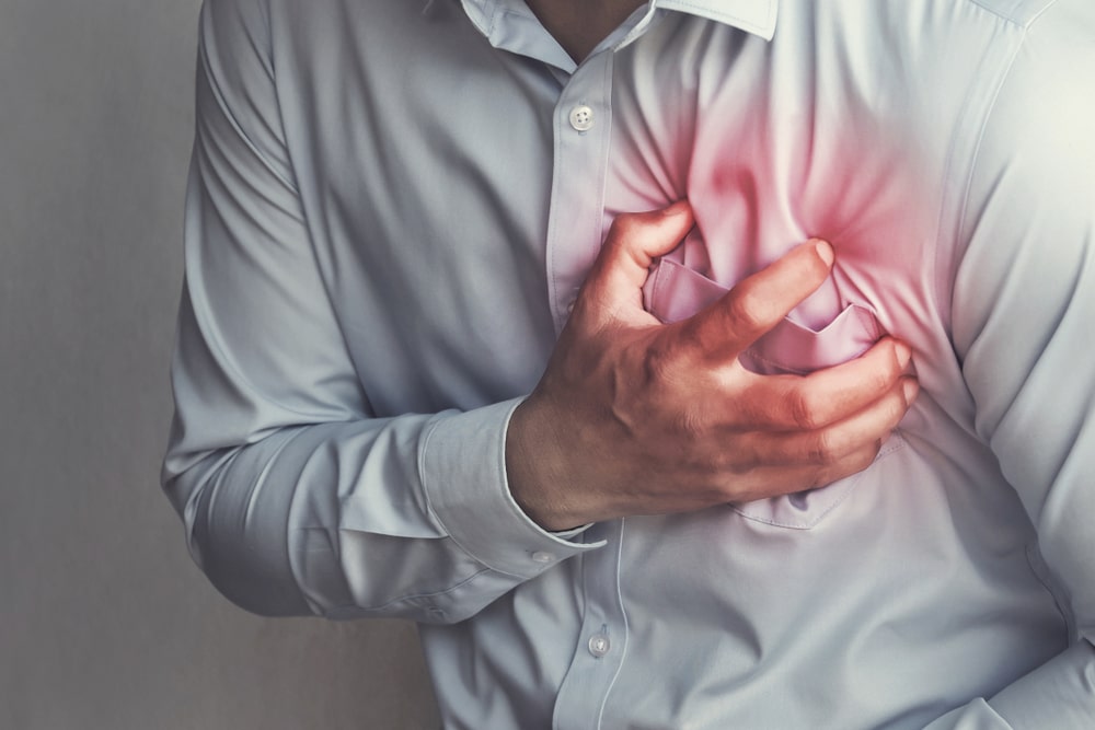
Typical chest pain in acute MI has the following characteristics:
- Intense and unremitting for 30-60 minutes.
- Substernal, and often radiates up to the neck, shoulder, and jaw, and down the left arm.
- Usually described as a substernal pressure sensation that also may be characterized as squeezing, aching, burning, or even sharp.
- In some patients, the symptom is epigastric, with a feeling of indigestion or fullness and gas. Other symptoms of myocardial infarction include the following:
- Anxiety, commonly described as a sense of impending doom.
- Pain or discomfort in areas of the body, including the arms, left shoulder, back, neck, jaw, or stomach.
- Light-headedness, with or without syncope.
- Cough.
- Nausea, with or without vomiting.
- Profuse sweating.
- Shortness of breath.
- Wheezing.
- Rapid or irregular heart rate.
- Fullness, indigestion, or choking feeling.
Treatment of Myocardial Infarction:
The primary goal of the treatment is to first open the blocked artery and restore blood flow to the heart muscle. This process is called reperfusion. As the reperfusion takes place, the patient is slowly relieved from the pain. The early reperfusion i.e. within 4-6 hours of the heart attack can not only prevent further damage to the heart but also preserves the pumping action of the heart. The damage to the pumping action of the heart develops heart failure, decreased ability to exercise, and abnormal heart rhythms.
Emergency agents:
Emergency agents are used in the process of reperfusion of the heart muscle. These agents help in relieving the severe heart pain, cause Vasodilatation to open the blocked artery, restore the oxygen supply and prevent further damage to the heart muscle. These agents are morphine, oxygen, nitroglycerine, aspirin.
Anti-platelet agents:
Anti-platelet medications prevent the formation of blood clots in the arteries. In NSAID, aspirin inhibits the cyclooxygenase-1 enzyme and thus prevents blood clotting by blocking the production of thromboxane A-2 by platelets, the chemical that causes platelets to clump. In addition to thromboxane A-2, platelets also produce adenosine diphosphate (ADP), which when acts on its receptor cause clumping of the platelets. The thienopyridines, clopidogrel, block the ADP receptor thus preventing the platelets from clumping.
Anti-coagulants:
Anti-coagulant medications prevent the growth of blood clots in the arteries. Anti-coagulants such as intravenous or subcutaneous heparin, subcutaneous low molecular weight heparin, and oral warfarin, prevent the formation of blood clots either by inhibiting the production of clotting factors or by interfering with the action of the clotting factors. e.g. Enoxaparin.
Clot-dissolving medications:
Fibrinolytic or thrombolytic agents are known as clot-dissolving agents used to open blocked arteries and dissolve the existing clots. Intravenous administration of clot-dissolving drugs such as tissue plasminogen activator (TPA) or TNK can open up to 80% of acutely blocked coronary arteries. Drugs such as streptokinase can be used for breaking up or dissolving the clots, by converting the intrinsic plasminogen present in the fibrin clot to its active agent form plasmin.
β-adrenergic receptor blockers:
β-blockers act by decreasing the workload of the heart. A decrease in the workload decreases the demand for oxygen by the heart and limits the amount of damage to the heart muscle. Long-term administration of β-blockers following a heart attack has been shown to improve survival and reduce the risk of future heart attacks. Esmolol, Propranolol, Atenolol, Timolol.
Make sure you also check our other amazing Article on : Angina Pectoris