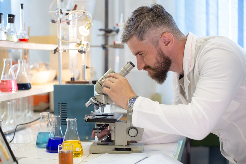
Parts of Compound Microscope: A microscope may be defined as ‘an optical instrument, consisting of a lens or a combination of lenses, for making enlarged or magnified images of minute objects’. Depending on the number of lenses, microscopes are classified into two types such as simple microscope and compound microscope, and depending on the number of the eyepiece, microscopes are classified into two types monocular microscope (single eyepiece) and binocular microscope (two eyepieces). Microscopes may be classified as light microscopes and electron microscopes, depending upon the source of illumination.
The compound microscope consists of three major systems.
(i) Support system: It comprises of base, stage, and body tube.
(ii) Illumination system: It throws light on the object for proper viewing. It comprises of light source or mirror, iris diaphragm, and condenser. The light source may be a concave or plain mirror or electrically illuminated by a tungsten filament lamp or a halogen lamp. Mirror and electric light sources are generally interchangeable.
(iii) Magnification system: It includes a set of lenses aligned in such a manner so that a magnified real image can be viewed. The objective is a set of lenses placed near the object. It partially magnifies the object, which can be observed through the eyepiece in a more magnified form.
Observation by a compound microscope, using the 100x objective requires cedarwood oil. It is popularly known as immersion oil and the lens is known as an oil immersion lens in microscopy. Immersion oil is colorless and has a refractive index similar to glass (1.55).
The refractive index of air is lower than that of glass and as light rays pass from the glass slide into the air, they are bent or refracted so that they do not pass into the objective lens. This would cause a loss of light which would reduce the numerical aperture and diminish the resolving power of the objective lens. Loss of refracted light can be compensated by using cedarwood oil, which has the same refractive index as glass, between the slide and the objective lens. In this way, decreased light refraction occurs and more light rays enter directly into the objective lens producing a vivid image with high resolution.
Parts of Compound Microscope
1. Oculars: A series of lenses (5x, 6x, 10x, 15x) that magnify the object and correct some of the defects of the objective. Huygenian, Ramsden, and compensating oculars are commonly used in microscopy.
2. Objectives: The objective is the most important lens on a microscope because its properties make the final image. The objective lenses generally equipped with a microscope are low power, high power, and oil immersion lens having a magnification of 10x, 40x, or 45x and 100x, respectively. The functions of the objective lens are to gather the light rays coming from any point of the object and unite the light at a point of the image and magnify the image. There are three types of objectives achromatic, fluorite, and apochromatic.
3. Condenser: This component is found directly under the stage and contains two sets of lenses that collect and concentrate light passing upward from the light source into the lens system. There are several different types of condensers, depending on the type of microscopy. e.g. Abbe condenser, variable-focus condenser, and achromatic condenser.
4. Iris diaphragm: It is equipped with a condenser. It controls the intensity of light entering the condenser. A lever is equipped with it to adjust the light intensity.
5. Illumination (light source): The light source is positioned at the base of the instrument. Some microscopes are equipped with a built-in light source to provide direct illumination. Others are provided with mirrors, with one side flat and the other concave. An external light source, such as a lamp, is placed in front of the mirror to direct the light upward into the lens system. The flat side of the mirror is used for artificial light and the concave side is for sunlight.
6. Body tube: Above the stage and attached to the arm of the microscope is the body tube. The upper end of the tube contains the ocular or eyepiece lens. The lower portion consists of a movable nose piece containing the objective lenses. It also provides sufficient space for image formation.
7. Revolving nose piece: A base in which the objectives are fixed and it holds 2 to 4 objectives and which can be revolved to align the required objective.
8. Focus adjustment knobs: There are two focus adjustment knobs, a coarse adjustment, and a fine adjustment. A coarse adjustment knob is used to bring the object into focus and a fine adjustment knob is used for fine and clear focus of the specimen.
9. Mechanical stage: It is a platform on which the specimen to be viewed is placed. Some stages have clips to hold the glass slide. Others have a mechanical stage, which makes it possible to move the slide across the stage.
Make sure you also check our other amazing Article on : Types of Cell Cultures
