Anatomy:
The kidneys lie on the posterior abdominal wall, one on each side of the vertebral column, behind the peritoneum, and below the diaphragms. The right kidney is usually lower than the left because of the considerable space occupied by the liver.
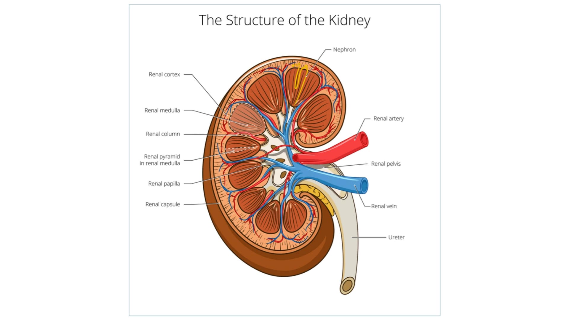
Kidneys are bean-shaped organs about 11cm long. 6cm wide, 3cm thick, and weigh around 150gms.
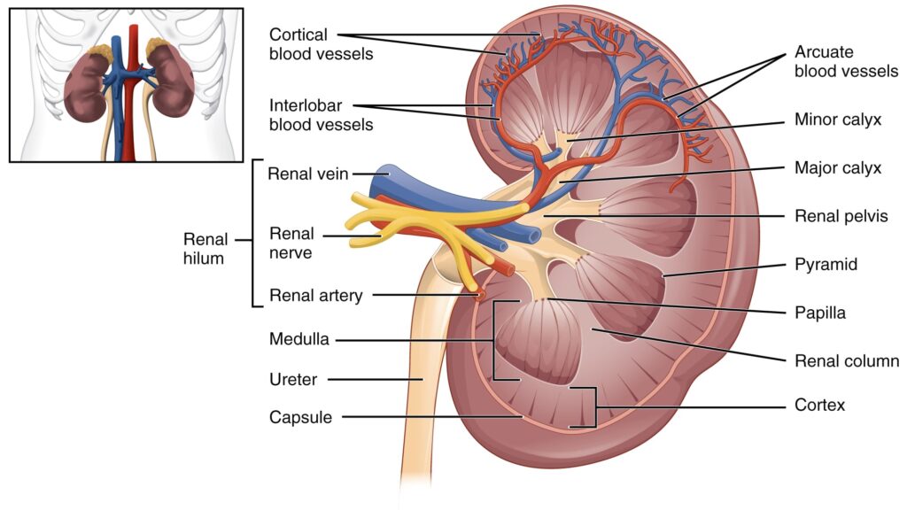
The kidney consists of a fibrous capsule surrounding the kidney, the cortex a reddish-brown layer of the tissue, and on the inner side, there are projections called pyramids and hollows called calyces.
Minor and major calyces unite form dilated part called the pelvis of the kidney
The pelvis terminates into a tubular structure called the ureter, which opens into the urinary bladder.
The urine formed in the kidney is transported to the urinary bladder through ureters. The urinary bladder passes the urine out through a tube called the urethra.
In males, the urethra is a common passage for urine and semen, in females, the urethra opens independently to the exterior.
The main functions of the kidney are as follows:
- Formation and secretion of urine
- Maintain the electrotele
- It maintains water equilibrium, pH equilibrium, osmotic equilibrium, and ionic equilibrium of the blood.
- Production and secretion of erythropoietin hormone that controls the formation of red blood cells.
- It excretes the waste products in dissolved form. These are the nitrogenous and sulfur-containing end products of protein metabolism.
- Production and secretion of renin an important enzyme in the control of BP
- It excretes poisonous and foreign substances from the body. This includes drugs, toxins, etc.
- It synthesizes new substances namely ammonia, hippuric acid, and inorganic phosphates.
Make sure you also check our other amazing Article on : Anatomy and Function of Liver